Social Media


Popular formats


Popular features
Photo

Filters

Stock royalty-free photos and images of Cellule
Discover unlimited high resolution images of Cellule and stock visuals for commercial use.


3D illustration of stem cell split into two. 3d stem cell. nucleolus, nucleus, nucleus of the eukaryotic cell. human body cell


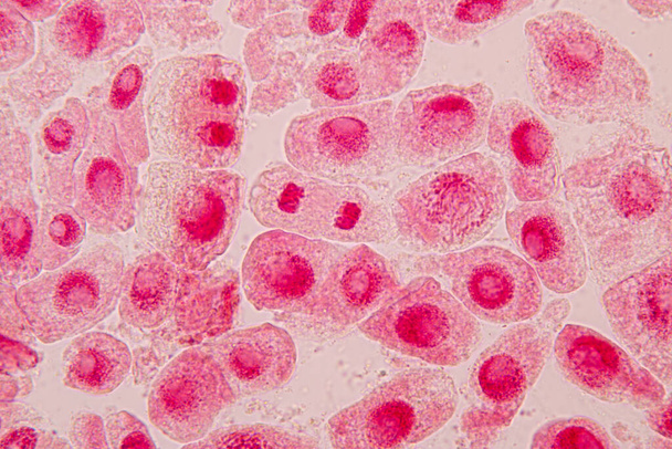
Prostate hyperplasia. Photomicrograph showing dilated glands, papillary projections inside the lumen of the glands, cystic dilatation with accumulation of secretory material


Chromosomes Human under the microscope for education.

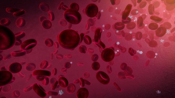
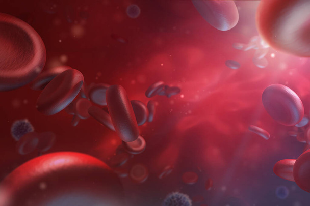
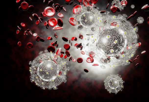
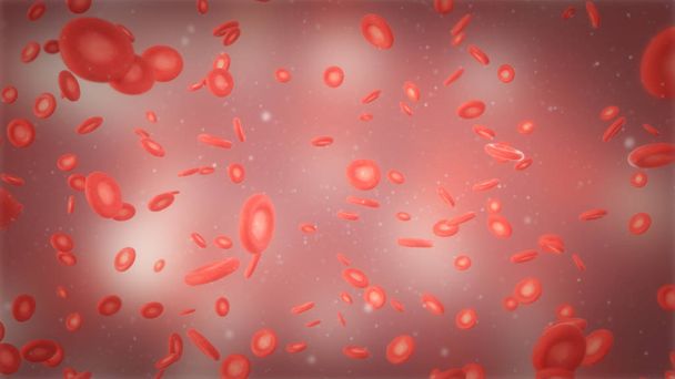
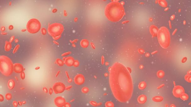
Red Blood Cells Flowing Through the Vein. Erythrocytes and leukocytes. Science and Health theme 3d illustration

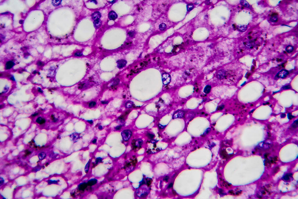
Histopathology of alcoholic hepatitis, light micrograph, photo under microscope. High magnification

3D illustration of 3d stem cell. nucleolus, nucleus, nucleus of the eukaryotic cell. human body cell.


Beautiful abstract blue bubbles in water extreme. Abstract nature pattrn for design. Macro photogrpaphy view.


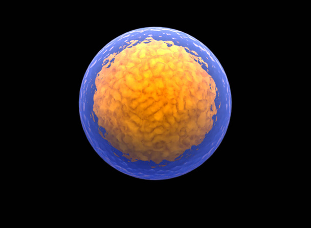
Orange spheres and molecular model, random distributed, 3d rendering. Computer digital drawing.


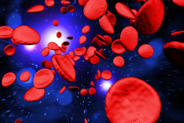
3D illustration of red blood cell

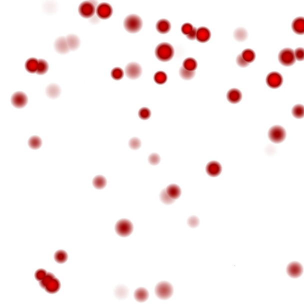
Red drops round isolated on white background


Bone marrow core biopsy pathology - disseminated Histoplasmosis


Human chromosomes under microscope view for education

Human chromosomes under microscope view for education


Abstract background of multiple bacteria.


Chronic pyelonephritis, light micrograph, photo under microscope

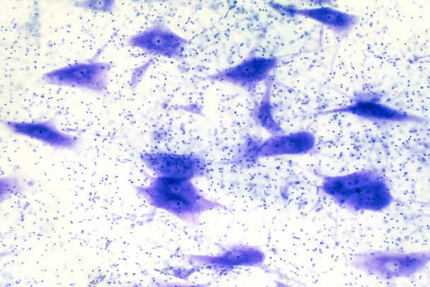
Neurons cells from the brain under the microscope view for education.

Strep throat also known as Streptococcus Pneumonia bacteria in mouth 3D digital illustration

Cell of a living organism, scientific concept. Illustration on a blue background. The structure of the cell at the molecular level, under a microscope. encrypted DNA in the cell, 3D illustration

Chromosomes Human under the microscope for education.

A man wearing a VR headset sits in a chair, deep in virtual conversation while holding a cell phone.

Histopathology of alcoholic hepatitis, light micrograph, photo under microscope. High magnification


T Cells attacking Cancer Cells- 3D illustration

Blood microscopic view. 3D medical illustration. Red and white blood cells. Illustration wallpaper render

Animal blood cells flowing through veins. Circulatory system search concept

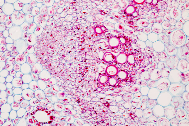
The study Plant tissue of under the microscope for classroom education.


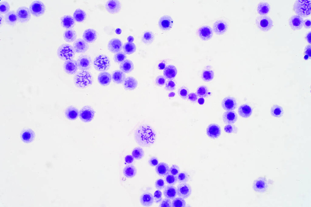
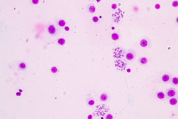
Blood cell formation from bone marrow.Acute lymphoblastic leukemia(ALL),fine with microscope(100x)


3D rendering of Beauty glossy bubbles in water,Abstract science background,Macro shot.

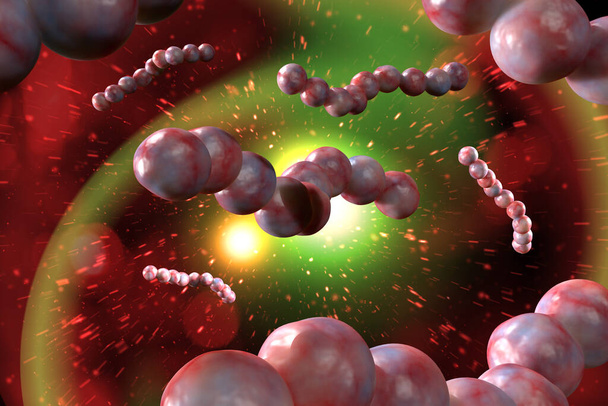
Shape of bacterial cell: cocci, bacilli, spirilla bacteria background

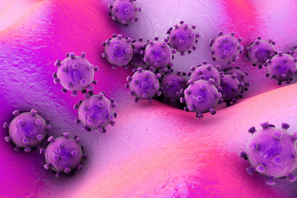
3D generated illustration of HIV Aids virus cells for medical science background

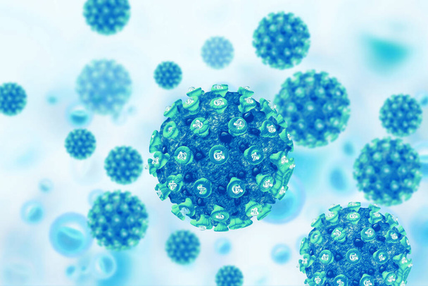
Virus cell on scientific background. 3d illustration

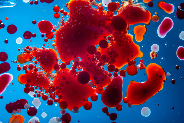
Red bubbles of blood cells on a blue background closeup. Abstraction of medicine and science. The concept of micro processes and diseases in the human body

3D illustration of red blood cell


3d rendering close up of microscopic cells flying in a spiral towards bright light. Abstract particles medical background. Blue tones.


A blood smear is often used as a follow-up test to abnormal results on a complete blood count (CBC) to evaluate the different types of blood cells.


Fake microorganisms. microbiology. Bacteria, cells, viruses, germs, microorganisms.


Prostate hyperplasia. Photomicrograph showing dilated glands, papillary projections inside the lumen of the glands, cystic dilatation with accumulation of secretory material


Virus destroys human dna chain. Genome structure. helix molecule in the body


Human chromosomes under microscope view for education

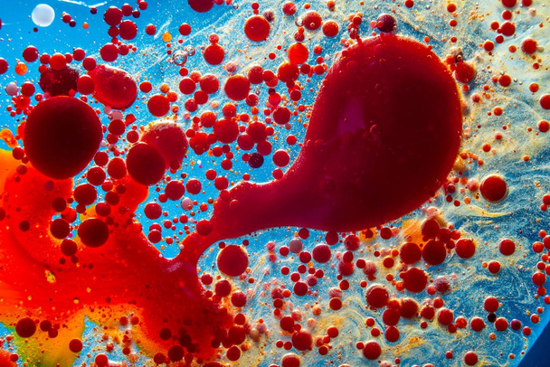
Red bubbles of blood cells on a blue background closeup. Abstraction of medicine and science. The concept of micro processes and diseases in the human body


Neurons and nervous system in the human brain. Histology of human brain tissue. Photo under microscope view.


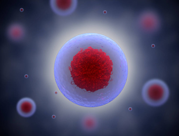
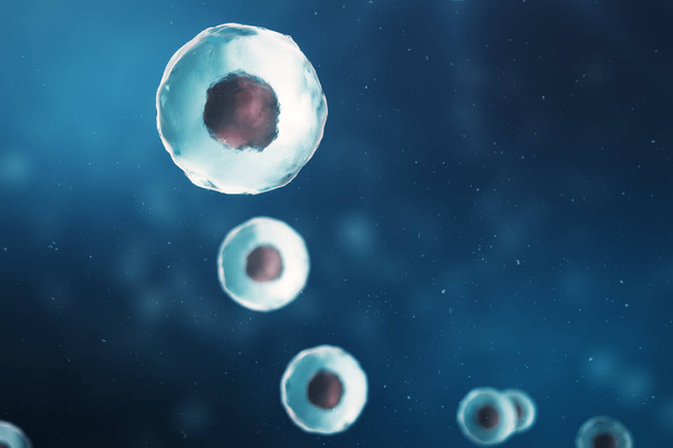
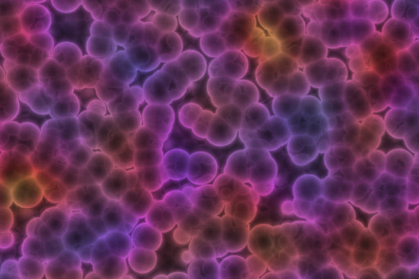
Human cells or animal. Cell colony. Concept of science and medicine, the regeneration of cells, the renewal of cells in the living organism, 3D illustration

3D illustration of red blood cell

3D illustration of 3d stem cell. nucleolus, nucleus, nucleus of the eukaryotic cell. human body cell.


Fine small structure formed by cells and lines


Crytpococcus neoformans stained with mucicarmine in lung tissue

Root tip of Onion and Mitosis cell in the Root tip of Onion under a microscope.

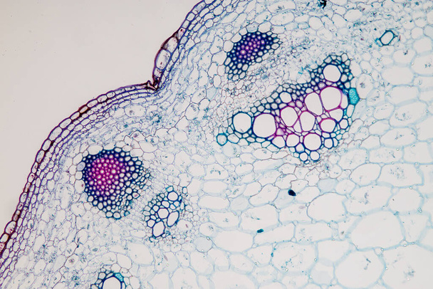
The study Plant tissue of under the microscope for classroom education.

Healthy human red bloodcells in close up 3d illustration

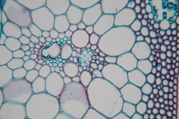
The study Plant tissue of under the microscope for classroom education.

Viruses on the surface of skin or mucous membrane, 3D illustration


Root tip of Onion and Mitosis cell in the Root tip of Onion under a microscope.