


Filters

Stock vector images of Membran
Discover royalty-free, professionally-designed vector art of Membran for personal and commercial use.

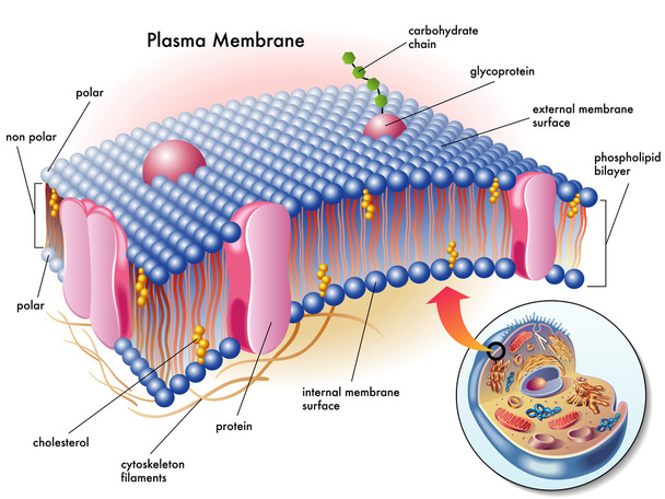
Medical illustration of elements of plasma membrane.
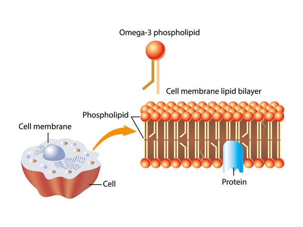
Omega 3 phospholipid cell membrane structure
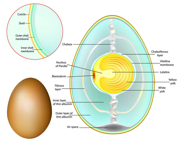
The structure of a chicken egg. Chicken Egg Development
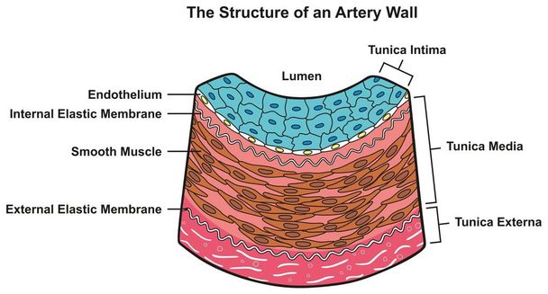
Structure of Artery Wall infographic diagram including all layers tunica externa media and intima cross section for medical science education and anatomy
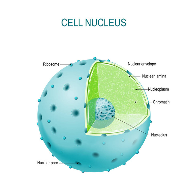
Structure of Nucleus. parts of the cell nucleus: nuclear envelope, nucleoplasm, nuclear matrix, chromatin and nucleolus

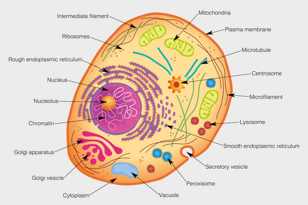
Realistic human cell anatomy diagram infographic poster vector illustration

Anatomy of animal cell with words illustration

Amoeba unicellular animal with pseudopods that lives in fresh or saltwater. Anatomy of an amoeba. Vector diagram.

Structure mitochondrion organelle found in most eukaryotic cells vector diagram

Labelled diagrams of typical animal and plant cells with editable layers.
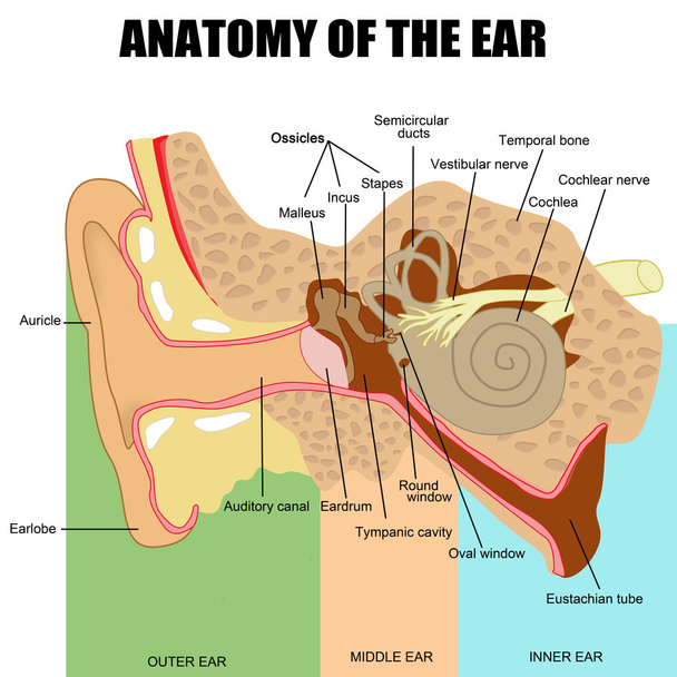
Anatomy of the human ear (useful for education in schools and clinics ) - vector illustration

Cell structure, cross section of the cell detailed anatomy with description

Structure of human cells. Organelles. The core nucleus, endoplasmic reticulum, Golgi apparatus, lysosomes, ribosomes, mitochondria, centriole Vector illustration on a black background
The graphic shows the elements of a human cell. Vector image

Cancer and cytotoxic T-cells. T lymphocyte kills cancer cells. T-cell (immune responses), release the perforin and granzymes, and attack cancerous cells. Through the action of perforin, granzymes enter the cytoplasm of the target cell, and lead to ap

Otitis externa ear disease 3d medical vector illustration on white background
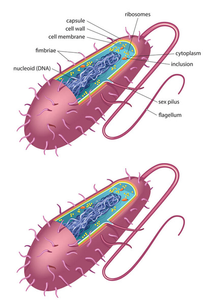
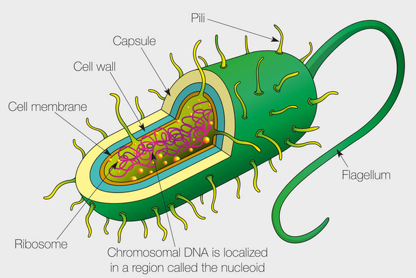
Illustration of typical bacterial cell - bacillus type

Mitosis is the process by which our bodies replace cells. Daughter cells have identical chromosomes to parent cell, genetic material remains constant. steps cell division. Vector diagram

Illustration of the human blood

The internal structure of an animal cell. Illustration

The structure of the ribosome. Functions. Infographics. Vector illustration on isolated background.
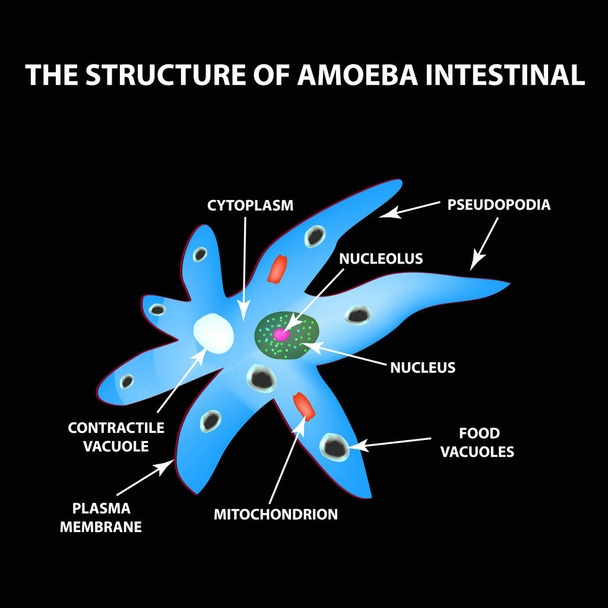
The structure of the amoeba is intestinal. Gastrointestinal Amebiasis. Infographics. Vector illustration on black background
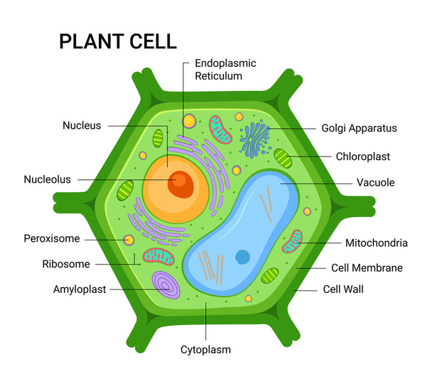
Vector infographic of the Plant cell anatomy structure. Nucleus, mitochondria, endoplasmic reticulum, golgi apparatus, cytoplasm, wall membrane etc
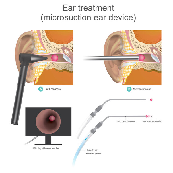
The Micro suction ear device it is vacuum working system
Human cells set of isolated icons with muscle fibers bone and stem cells diagram with text vector illustration

Cell membrane proteins. Phospholipid bilayers structure of cytoplasmic membrane

The graphic shows the elements of the nucleus of a human cell with their names. Vector image

Structure of Bacterial Cell. Cutaway vector diagram of a typical bacterial cell illustrating structural components

Human cell membrane structure illustration

Ear anatomy 3d medical vector illustration isolated on white background
The graphic shows the parts of a bacterium cell. Vector image

Lysosome anatomy. Hydrolytic enzymes, Membrane and transport proteins. This organelle use the enzymes to break-down virus particles or bacteria in phagocytosis of macrophages. Vector illustration

Realistic blood vessels artery and vein composition with the structure of blood vessels headline and descriptions vector illustration
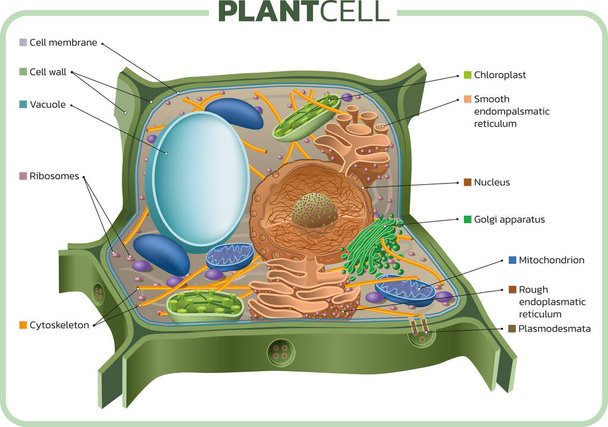
Vector illustration of the basic structure of the plant cell.

Effect of different solutions on blood cells.The effect of osmosis on cells. Hypotonic, Isotonic, and Hypertonic solution. Tonicity
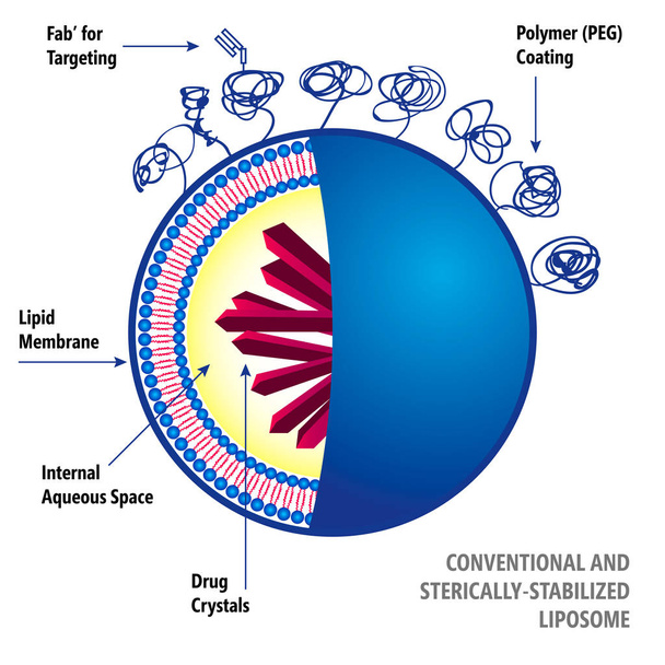
Medical vector illustration of liposomes drug delivery system

Cartoon ear anatomy. Human sound sensory organ medicine infographic, ears internal structure vector illustration. Ear infographic anatomy. Ear health organ medical, sensory biology
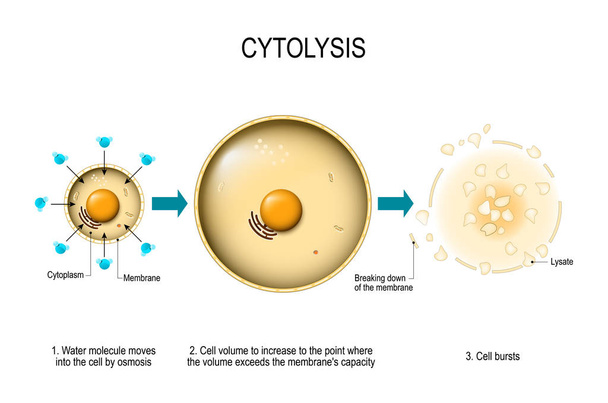
Cytolysis. Osmotic lysis. Water enter the cell and causes its volume to increase to the point where the volume exceeds the membrane's capacity and the cell bursts. Vector diagram for educational, medical, biological and science use
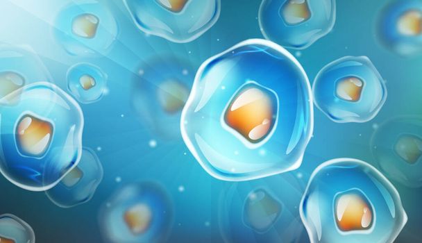
Cells under a microscope. Research of stem cells. Cellular Therapy. Cell division. vector illustration on a light background.

Ear anatomy diagram,vector illustration.

Anatomy of a Bird Embryo illustration

Detailed Diagram Models of a Cell Membrane

Anatomy of the Lysosome: Hydrolytic enzymes, Membrane and transport proteins. This organelle use the enzymes to break down and digest food particles, engulfed viruses or bacteria in the cell. Vector diagram for medical use

Vector illustration of Examination ear anatomy
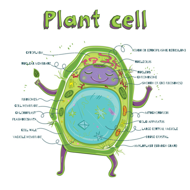
Cartoon vector illustration of structure of plant cell. Illustration showing the plant cell anatomy
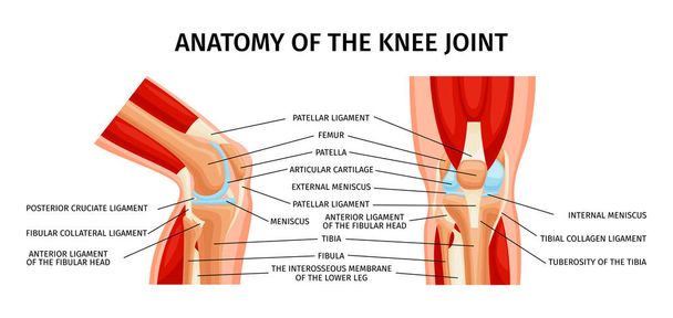
Healthy knee joint anatomy front and side view diagram realistic infographics with labelled parts vector illustration
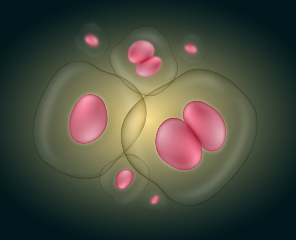
Group of cells over a green background

Prokaryotic cell structure diagram vector

Meninges anatomy structure part infographic diagram human body central nervous system layers including dura archnoid pia mater skull brain neurology biology physiology science education vector

Vector illustration with human human respiratory system affected by the coronavirus
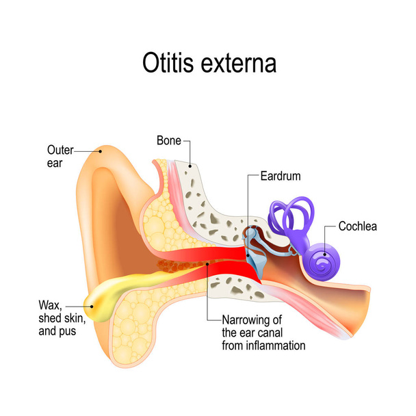
Swimmer's ear. Otitis externa is inflammation of the ear canal. Human anatomy. Vector illustration for medical use

Vector illustration of an animal cell. cell wall structure
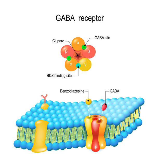
Cell membrane. GABA receptor and various sita for ligands bind. Top view of ion channel which illustrates the five combined subunits that form Cl ion channel pore.

Cartoon vector illustration of structure of plant cell. Illustration showing the plant cell anatomy

Eukaryote cell vector simple illustration

Shoulder Joint of Human Body Anatomy infographic diagram with all parts including bones ligaments muscles bursa cavity capsule cartilage membrane for medical science education and health care

GABA receptor. ligand-gated ion channel, metabotropic receptors. Neurotransmitter in the central nervous system.

Micro bacterium or virus organisms. Microscopic lactobacillus or acidophilus organism abstract background with bokeh blur defocused elements. Illustrated vector
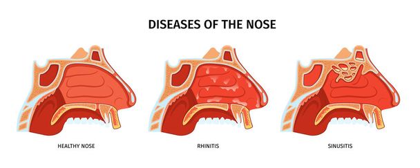
Diseases of nose anatomical cross section with healthy nose sinusitis and rhinitis realistic vector illustration
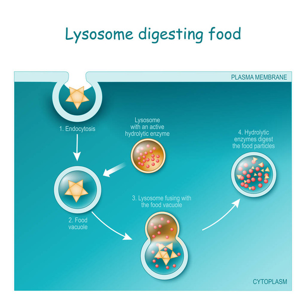
Endocytosis. Lysosome digesting food. Part of cell (plasma membrane, cytoplasm and lysosome), with food vacuole. Lysosome fusing with the food vacuole. Vector illustration

Schematic chicken egg anatomy stock vector illustration, Is marked thin albumen, chalaza, yolk, vitelline membrane, germinal disk, eggshell, cuticula, air cell, thick albumen

Anatomy of the Ear. Illustration
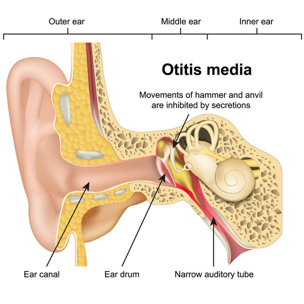
Otitis media ear disease 3d medical vector illustration on white background

Amoeba anatomy. unicellular animal with pseudopods. Vector illustration for medical, educational and science use

Vector bacterial cell anatomy isolated on white background. Educational illustration.
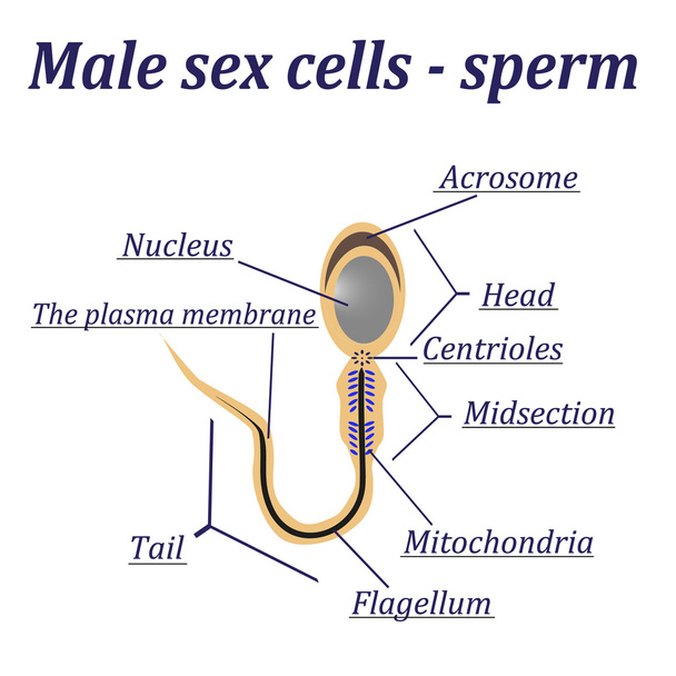
Diagram of the male sex cells - sperm. Isolated on a white background.
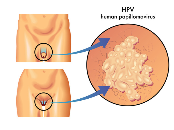
Vector illustration of colorful Papillomavirus scheme

Human cell anatomy realistic infographics with labelled educational diagram on white background vector illustration

Coronavirus cell structure concept /illustration of 2020 Novel coronavirus showing single-stranded RNA genome and spike, membrane and envelope proteins of the virus.

Animal Cell Color Diagram of organelles inside the cell membrane for science and biology concepts.
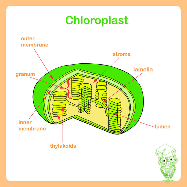
Scheme of Chloroplast structure, hand drawn biology stock vector illustration

Vector illustration of l-carnitine transports fat molecules into the mitochondria.

Babies of three-parent. inherited mitochondrial diseases. Pronuclear transfer in human embryos. Vector diagram for educational, medical, biological and science use

Plant cell anatomy structure. Close-up of Organelles of a plant cell. Educational infographic. Vector illustration

Anatomy of the human thyroid

Ruptured eardrum or perforated eardrum. Tympanic membrane perforation. Hole in the eardrum.
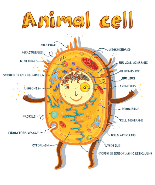
Cartoon vector illustration of structure of animal cell. Illustration showing the animal cell anatomy

Endocytosis. phagocytosis is cell eating, pinocytosis is a cell drinking, receptor-mediated endocytosis - when cells absorb metabolites, hormones, proteins and viruses by receptors on the surface of the cell.

The structure of the Golgi apparatus. Infographics. Vector illustration on isolated background.
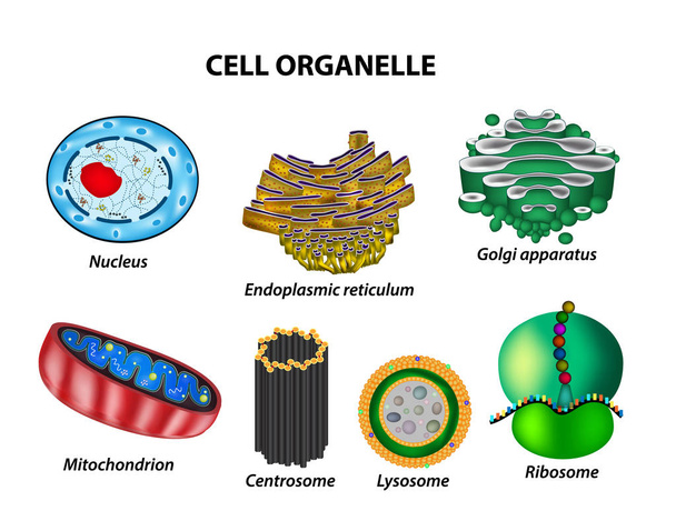
Set the cell organelles. Nucleus, endoplasmic reticulum, Golgi apparatus, mitochondria, centrosome, lysosome, the ribosome. Infographics. Vector illustration on isolated background.

Binary fission in amoeba. Vector educational illustration. Reproduction
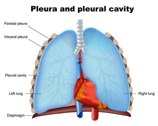
Lung pleura and pleural cavity medical vector illustration on white background

Anatomy of plant cell (Biology Diagram) illustration
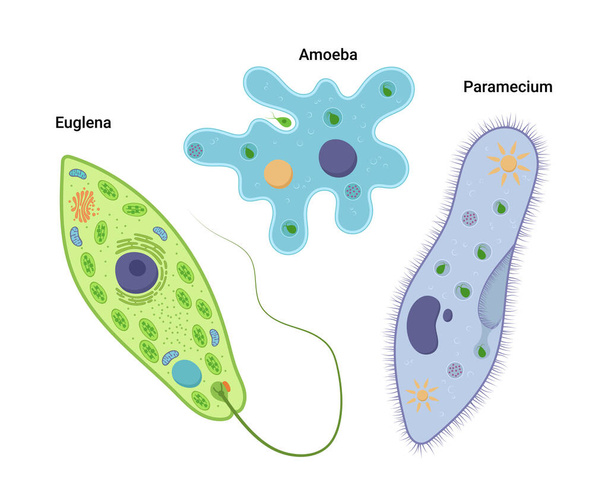
Vector illustration of unicellular organisms. Amoeba proteus Paramecium caudatum and Euglena viridis. Protozoa
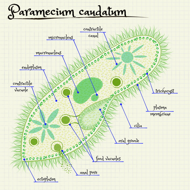
Vector drawing of the structure of Paramecium saudatum
Human cells flat set of isolated icons with colorful images of microorganisms and internal bacteria shapes vector illustration

Group of cells over a green background
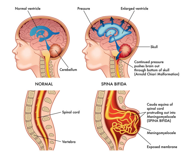
Medical illustration shows a child in two situations, left healthy and right afflicted by spina bifida with annotations explaining the symptoms of the pathology.

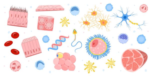
Different biology cells on a white background

Chart showing plasma membrane illustration

Vector drawing of the structure of a single-celled fungus
Cell-division vector icon. Modern vector illustration concepts. Easy to edit and customize.

Endocytosis. Process of vesicle transport for endocytosis

Illustration of the anatomy of the plant cell
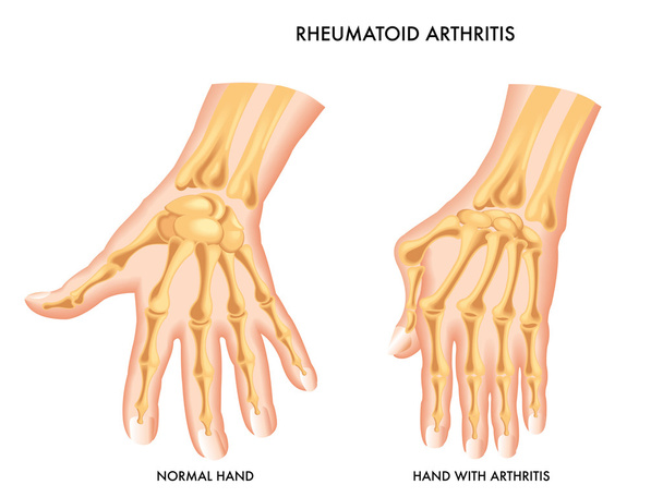
Rheumatoid Arthritis
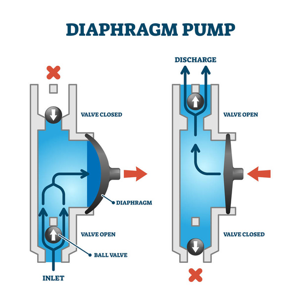
Diaphragm or membrane pump working process example, technical diagram drawing with fluid flow principle. How it works labeled visual example vector illustration. Cross section with water chamber.
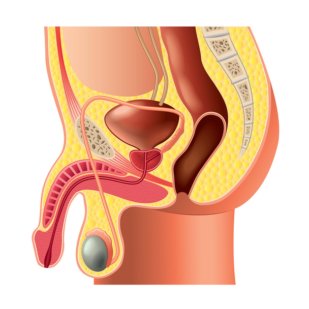
Male reproductive system anatomy isolated photo-realistic vector

Chloroplast structure. Plant cell, chloroplast, granum and thylakoid.
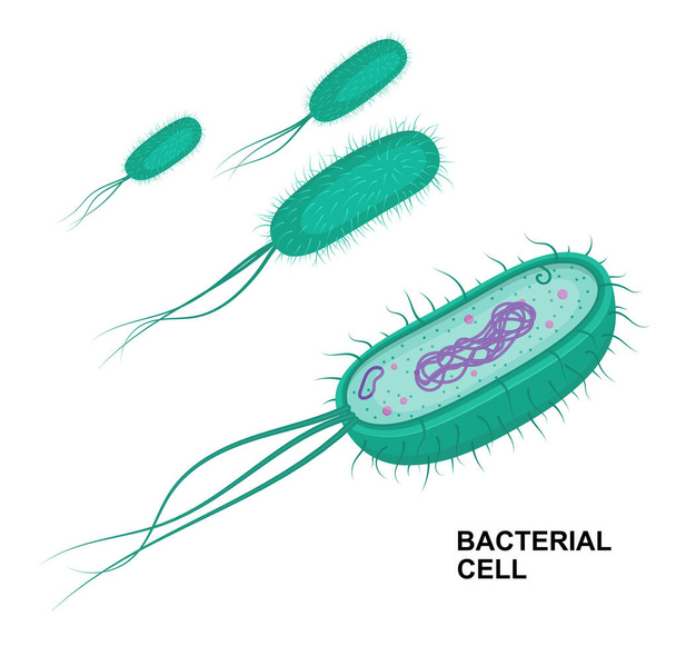
Vector bacterial cell anatomy isolated on white background. Educational illustration. Structure of prokaryotic