Social Media


Popular features
Photo

Filters

Stock royalty-free photos and images of Клітини
Discover unlimited high resolution images of Клітини and stock visuals for commercial use.
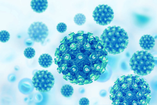
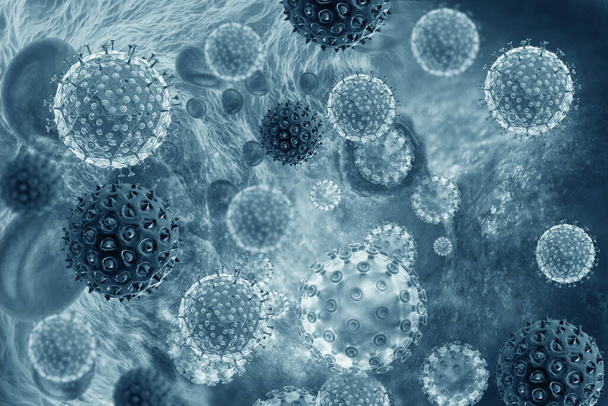
Virus cell on scientific background. 3d illustration

Renal cell carcinoma, light micrograph, photo under microscope

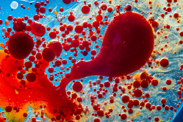
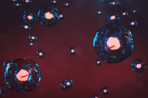
Liquid Bubble Abstract Design Background

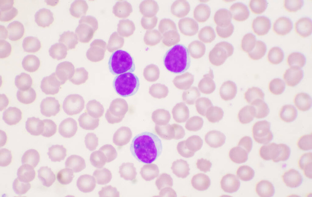
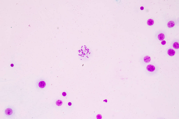
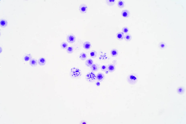
A blood smear is often used as a follow-up test to abnormal results on a complete blood count (CBC) to evaluate the different types of blood cells.Chronic lymphocytic Leukemia(CLL)

Bubbles red oil, serum look like a nucleus isolated on black background.
Virus cell on scientific background. 3d illustration

Fake microorganisms. microbiology. Bacteria, cells, viruses, germs, microorganisms.

Human chromosomes under microscope view for education
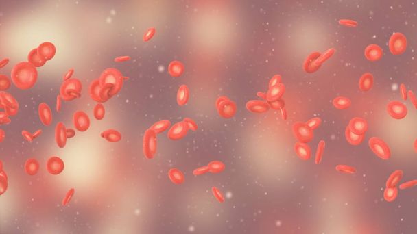
Red bubbles of blood cells on a blue background closeup. Abstraction of medicine and science. The concept of micro processes and diseases in the human body

Liquid Bubble Abstract Design Background

Orange spheres and molecular model, random distributed, 3d rendering. Computer digital drawing.

Chromosomes Human under the microscope for education.

Bright neon favia with green centers and a blue base

Close up beautiful abstract blue bubbles in water extreme. Abstract nature pattrn for design.

Fine small structure formed by cells and lines

Blood cells and viruses, biology concept background
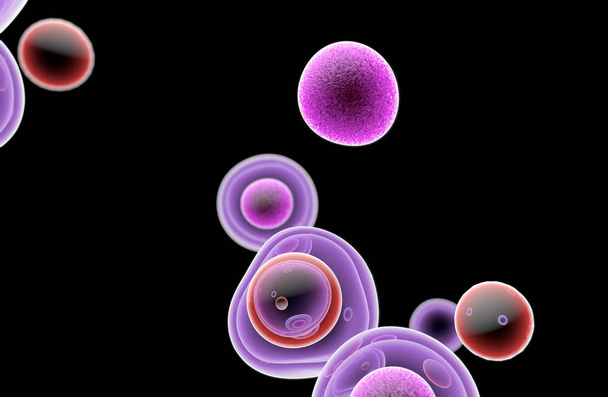
Embryonic Stem Cells Cellular-Therapy Regeneration

Close up Beautiful abstract soap bubbles. Background pattern for design. Macro photography view.
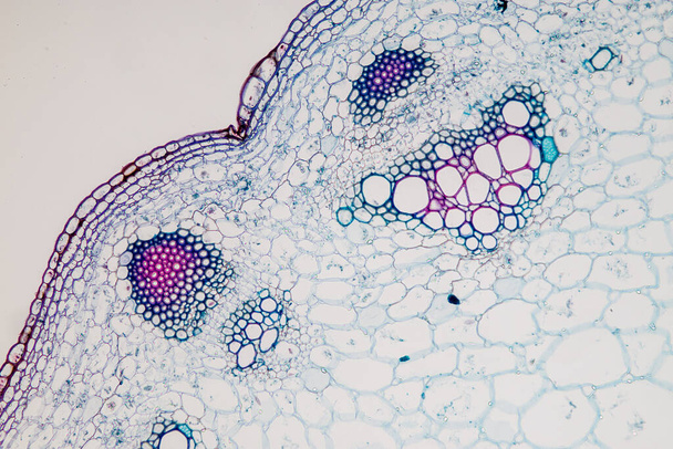
The study Plant tissue of under the microscope for classroom education.

Red blood cells (red blood cells) under the microscope, 3D rendering

White blood cells in medical background concept.

Red coronavirus microscope,bacteria, disease, microorganisms. close-up copy space

Virus destroys human dna chain. Genome structure. helix molecule in the body

Beautiful abstract pink bubbles background pattern for design. Liquid fluid texture for background, banner.
Cell of a living organism, scientific concept. Illustration on a blue background. The structure of the cell at the molecular level, under a microscope. encrypted DNA in the cell, 3D illustration

Histopathology of prostate gland hyperplasia, light micrograph, photo under microscope

Human chromosomes under microscope view for education

Image of bacteria in laboratory placed in a receptacle
White blood cell Mature lymphocyte on red blood cells background.
Virus seamless generated hires texture

Macro of oil drops and pigment on water surface with bright background

Graphic conceptual illustration of Coronavirus cells background header. Colorful, abstract design in soft focus uses paint spatters and brush strokes details.
Virus seamless generated hires texture

Shape of bacterial cell: cocci, bacilli, spirilla bacteria background

Coronavirus 2019-nCoV 3D rendering model. Microscopic view of a infectious virus and red blood cell in the human blood vessels.

Red blood cells on dark red background. 3D render

Human chromosomes from blood under microscope view for education.

Red and white blood cells floating through vein

Human lung tissue under microscope view. Lungs are the primary organs of the respiratory system in humans and many other animals

Monocytes are a type of white blood cells (leukocytes).

Realistic molecule bacterium illustration graphics 3d microbe

3D rendering of microorganisms, viruses, and red blood cells in a cell. Illustration for medical compositions, the idea of health protection, protection from coronavirus. Medical background.

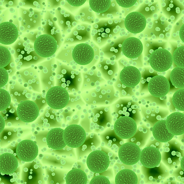
Green single cell chlorella algae microscopic conceptual 3D illustration collage
Chromosomes Human under the microscope for education.

Smartphone on whiteslim side view

A blood smear is often used as a follow-up test to abnormal results on a complete blood count (CBC) to evaluate the different types of blood cells.

Lewy bodies in parkinson's disease (PD) or dementia (LBD) - isometric 3d illustration

3D illustration cell of a living organism, scientific concept. Illustration on a blue background. The structure of the cell at the molecular level, under a microscope. encrypted DNA in the cell
Coronavirus cells COVID-19 Infectious disease. Fast transmission spread disease. High concentration of coronavirus . 3D rendering 3D illustration

A blood smear is often used as a follow-up test to abnormal results on a complete blood count (CBC) to evaluate the different types of blood cells.

Shape of bacterial cell: cocci, bacilli, spirilla bacteria background

Microscope shot of abstract cell element on dark background

Virus on white background.3d render
White blood cell lymphocyte on red blood cells background hematology concept.

Root tip of Onion and Mitosis cell in the Root tip of Onion under a microscope.
Abstract bacteria bokeh seamless generated hires texture

Human lung tissue under microscope view. Lungs are the primary organs of the respiratory system in humans and many other animals

Micro probiotic microorganism, science background.

3d illustration water molecules and bubbles. H2o

Orange spheres and molecular model, random distributed, 3d rendering. Computer digital drawing.

The study Plant tissue of under the microscope for classroom education.

Characteristics of fungi living in wood as a group, are polyphyletic under the microscope for education.

3D generated illustration of HIV Aids virus cells for medical science background

Acrylic, paint, abstract. Closeup of the painting. Colorful abstract painting background. Highly-textured oil paint. High quality details.

Digital illustration of dna structure in 3d on digital background
Blood Cells with nucleus on scientific background.3d illustration

Microscope shot of abstract cell element on white background

Chronic pyelonephritis, light micrograph, photo under microscope. High magnification

Human chromosomes under microscope view for education
Human chromosomes under microscope view for education

Cellular structure, biology concept, 3d rendering. Computer digital drawing.

Macrophages infected by Leishmania amastigotes, isolated on white background, 3D illustration

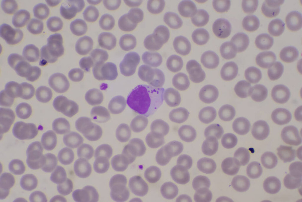
A lymphocyte is any of three subtypes of white blood cell in a vertebrate's immune system. All three are agranulocytes.
3D illustration of red blood cell
Abstract DNA research red Blood cells and stem cell human cell in blood vessels,microscope view ,health care concept cardiovascular diseases and hypertension and medical,3D rendering illustration

Adequate of platelet in blood smear compleat blood count.

Characteristics of Rhizopus is a genus of common saprophytic fungi on Slide under the microscope for education.

Lots of dissociative blue cells, 3d rendering. Computer digital drawing.

Virus infected blood cells. 3d illustration

Abstract molecular structure

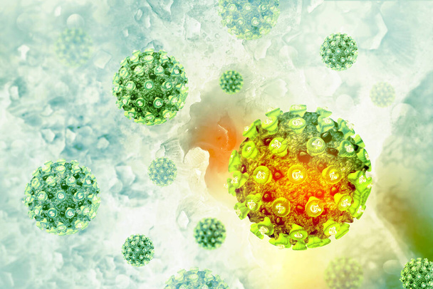
Deadly epidemic coronavirus cell closeup concept 3D Illustration

Cells microbiology molecules concept, 3D illustration

Stem cells on colorful background, 3D illustration

Coronavirus cells COVID-19 Infectious disease. Fast transmission spread disease. High concentration of coronavirus animation. 3D rendering 3D illustration
The cell floating in a human veins

Full frame pastel colored blurry abstract back for various purposes
Potato cells with starch corns under the microscope and in polarized light.

Nerve cell under the microscope - Abstract blue dots on white background.

360-degree spherical panorama of eosinophil, a white blood cell, in blood, 3D illustration. Eosinophils are granulocytes taking part in allergy and asthma, protection against multicellular parasites

Adequate of platelet in blood smear compleat blood count.

Blood smear is often used as a follow-up test to abnormal results on a complete blood count (CBC) to evaluate the different types of blood cells.Medical science background showing blast cells(AML)

Sepsis or septicaemia is a life-threatening illness.

Virus cell on scientific background. 3d illustration

A blood smear is often used as a follow-up test to abnormal results on a complete blood count (CBC) to evaluate the different types of blood cells.

3d render of Corona virus that causing the infection of MERS, influenza and SARS that outbreak around the world. COVID-19 virus that pandemic medical risk for human lung.

Abnormal neutrophil in blood smear.Sepsis or septicaemia is a life-threatening illness. Presence of numerous bacteria in the blood, causes the body to respond in organ dysfunction.

Coronavirus cells. Group of viruses that cause respiratory infections under the microscope. 3D rendering 3D illustration 3D illustration

Human Cells under microscope

Microscope photo of a sambucus stem with parenchyma cells.
Coronavirus 2019-nCov or Covid-19 concept. The virus attack to many people in the world and many people was die. The doctor tried to protected suspected case and controlled social distancing.