


Filters

Stock royalty-free photos and images of Lantio
Discover unlimited high resolution images of Lantio and stock visuals for commercial use.


Hand drawn medical illustration drawing with imitation of lithography: Pelvis 2 angels (male at the top and female at the bottom of drawing)

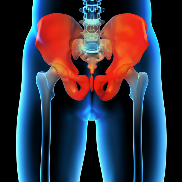
Anatomy of the pelvis bones, including the ilium, ischium, sacrum, and pubis, photorealistic 3D illustration. Front view. Perfect for educational or medical purposes.


Hip replacement model. 3D illustration

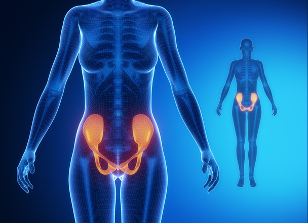
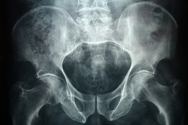
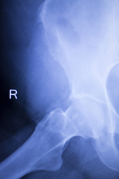
PELVIS blue x--ray bone scan


Illustration of human pelvis


3d rendered illustration of the female femur bone - front view

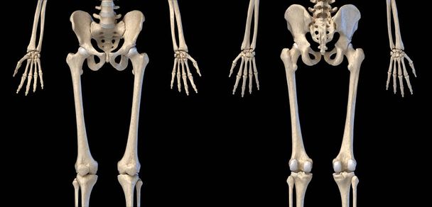
Human Anatomy, hip, limbs and hands skeletal system. Front and back views. On black background. 3d illustration.

Human Anatomy, hip, limbs and hands skeletal system. Front view. On black background. 3d illustration.

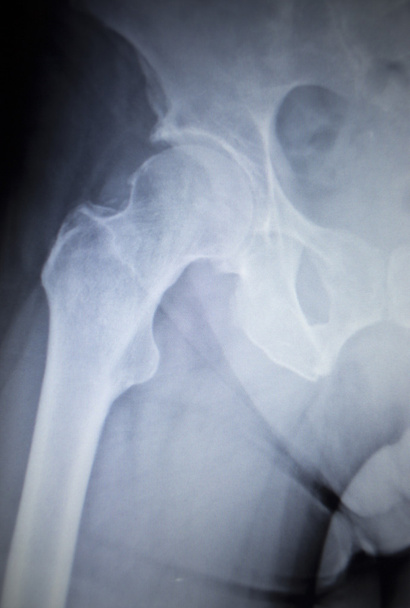
Hips pelvis xray scan test result.
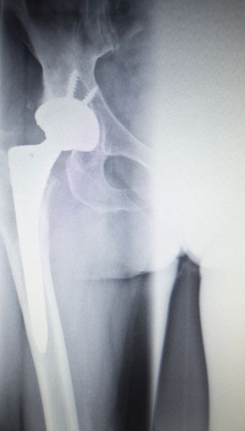
X-ray of the hip prosthesis elderly man
Scanning of an anterior posterior radiograph of the pelvis taken, among others radiographs, to try to detect the origin of pain in the hip of an adult man

Old x-ray picture of one deformed hip

The structure of the bones of the human legs. Sketch for artists. Anatomical diagram.
Negative X-Ray of spinal column, chest, abdomen, pelvis and thighbone of a female 16 years old small dog
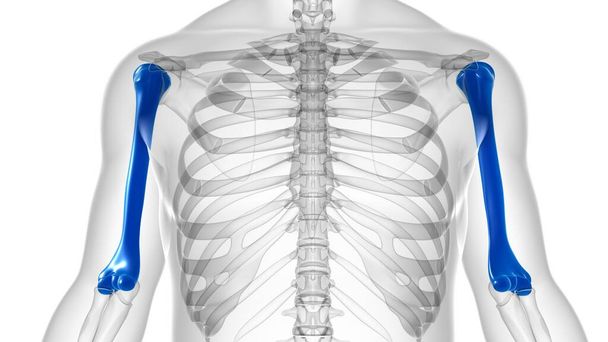
Human skeleton anatomy Humerus Bone 3D Rendering For Medical Concept

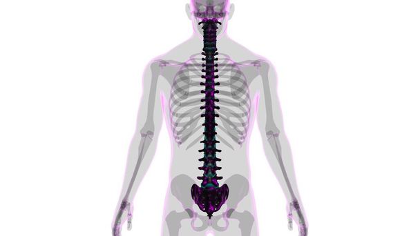
Human Skeleton Vertebral Column Lumbar Vertebrae Anatomy 3D Illustration

The bony skeleton is divided into 2 parts axial skeleton and appendicular skeleton 3D illustration

Man with PELVIS bones anatomy x-ray scan

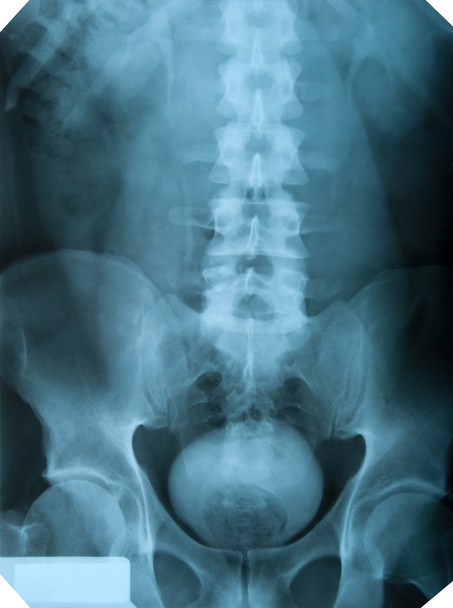
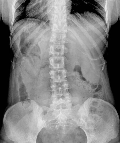
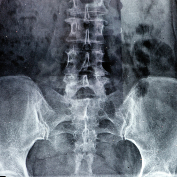
X-ray of the pelvis and spinal column.
Hip replacement xray orthopedic medical x-ray Traumatology test scan image of old age senior adult.
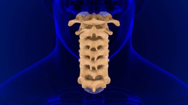
Human Skeleton Vertebral Column Cervical Vertebrae Anatomy 3D Illustration

Human Skeleton Vertebral Column Lumbar Vertebrae Anatomy 3D Illustration
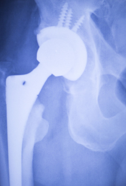
X-ray scan image of hip joints with orthopedic hip joint replacement implant head and screws in human skeleton in blue gray tones. Scanned in orthopedics traumatology surgery hospital clinic.

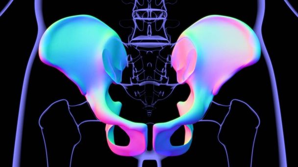
3D Illustration of Human Hip and Pelvis

3D reconstruction from patient images, 3D printed pelvis.

X-ray of the spine and pelvis. Medicine
Human Skeleton Hip or Pelvic bone Anatomy For Medical Concept 3D Illustration

Human skeleton anatomy Femur Bone 3D Rendering For Medical Concept
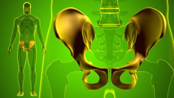
Human Skeleton Hip or Pelvic bone Anatomy For Medical Concept 3D Illustration
Human Skeleton Vertebral Column Vertebrae Anatomy 3D Illustration

Human skeleton anatomy Radius Bone 3D Rendering For Medical Concept

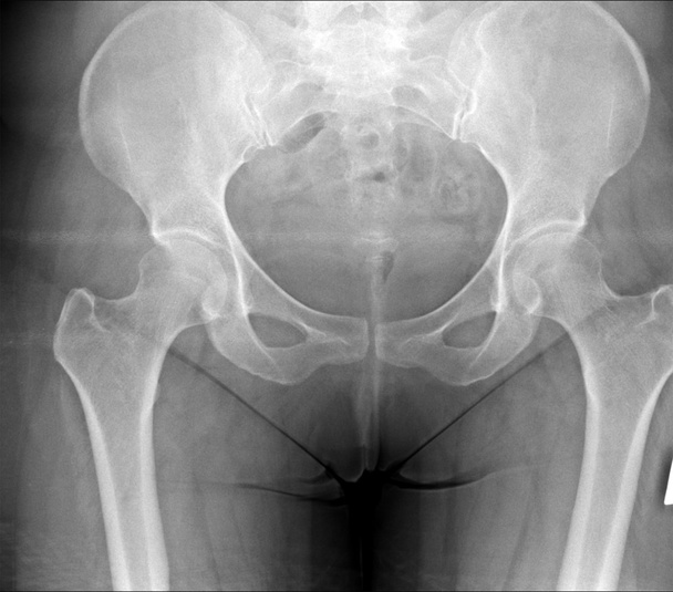
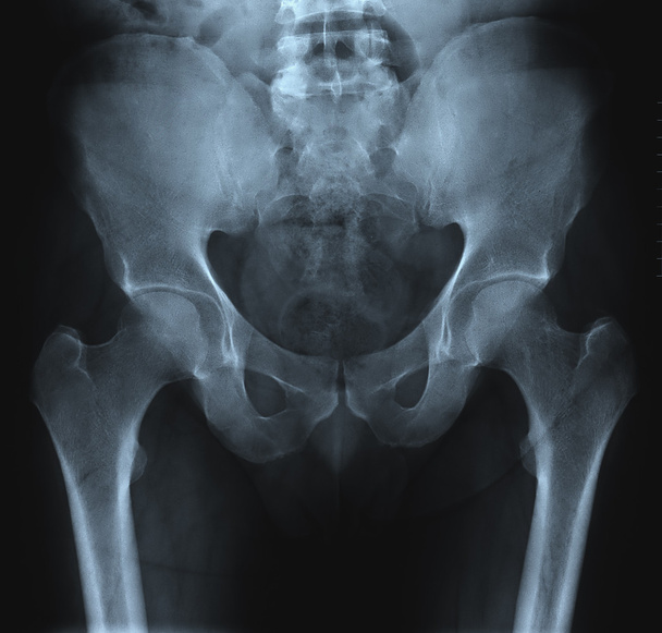
Radiograph of the pelvis for a medical diagnosis
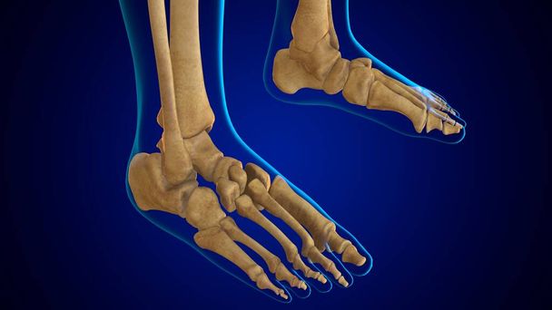
3D Illustration Human Skeleton Foot bone Anatomy

Articular capsule Anatomy For Medical Concept 3D Illustration

Human Skeleton Vertebral Column Vertebrae Anatomy 3D Illustration

Diagnostic shoulder and arm X-ray film of a patient.

X-Ray image hip medical concept

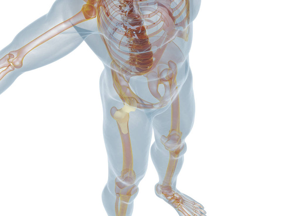
Fracture of the femoral neck on white background - 3D Rendering

Human Skeleton Foot bones Anatomy For Medical Concept 3D Illustration

Close up MRI. Whole spine intradural extramedullary mass at Lt lateral cord compression
3d rendered, medical illustration of a painful sacrum

Human Skeleton Hip or Pelvic bone Anatomy For Medical Concept 3D Illustration
Hip replacement model. 3D illustration

Human skeleton anatomy Femur Bone 3D Rendering For Medical Concept

X-ray photography of the human lumbo-sacral pelvis and spinal column of adult people

Hamstring muscle group anatomy model. 3d illustration.

The bony skeleton is divided into 2 parts axial skeleton and appendicular skeleton 3D illustration

X-ray of the spinal column and pelvis of a woman

Hip replacement xray orthopedic medical x-ray Traumatology test scan image of old age senior adult.
Pelvis frontal xray image for medical diagnosis

Hip, leg, thigh and pelvis injury medical x-ray test scan image of old age senior adult.
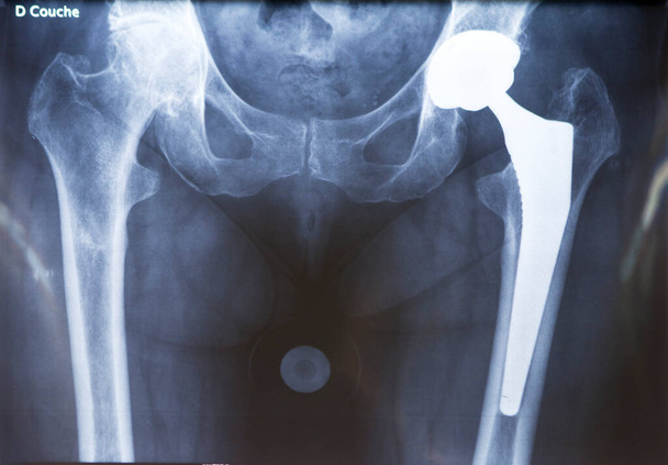
X-ray of a hip prosthesis.
Hip replacement xray orthopedic medical x-ray Traumatology test scan image of old age senior adult.

X-ray scan image of hip joints with orthopedic hip joint replacement implant head and screws in human skeleton in blue gray tones. Scanned in orthopedics traumatology surgery hospital clinic.
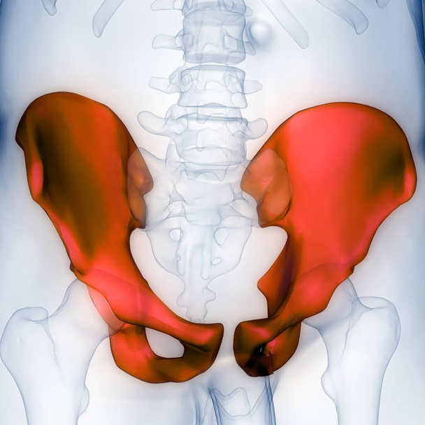
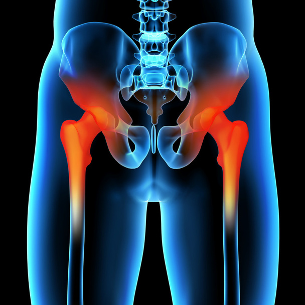
Human Body Bone Joint Pains (Hip and Pelvis Anatomy). 3D

Human Skeleton Hip or Pelvic bone Anatomy For Medical Concept 3D Illustration
Xray of a male pelvic, it's a high resolution 24 mp photo not a scan.
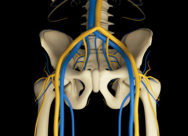
Human pelvis anatomy model. 3d illustration

Pelvis, Human skeleton, Female Pelvic Bone anatomy, hip, 3D artwork, Bones Labeled Anatomy top View, black background

Hip replacement xray orthopedic medical x-ray Traumatology test scan image of old age senior adult.
FIBULA black x--ray bone scan
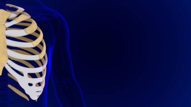
Human skeleton anatomy Rib Cage 3D Rendering For Medical Concept

X-ray film

Human bones joints and ligaments Anatomy For Medical Concept 3D Illustration

Medically 3D illustration showing right kidney, x-ray style, cross section
Hip, leg, thigh and pelvis injury medical x-ray test scan image of old age senior adult.
3d rendered, medical illustration of a painful sacrum
X-ray scan image of hip joints with orthopedic hip joint replacement implant head and screws in human skeleton in blue gray tones. Scanned in orthopedics traumatology surgery hospital clinic.

Joint pain can be caused by injury affecting any of the ligaments, bursae, or tendons surrounding the joint.

Human Skeleton Skull Mandible Bone Anatomy For Medical Concept 3D Illustration

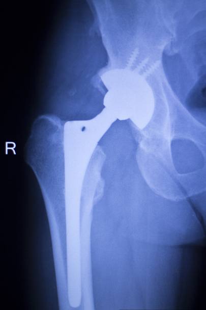
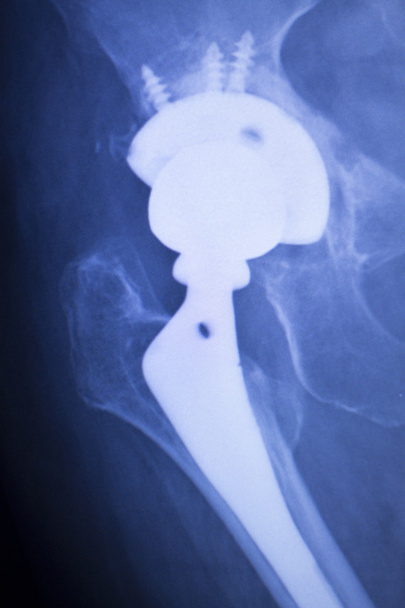
Hip joint replacement xray showing ball and socket joint's titanium screw implant in medical orthpodedics scan.

Running woman with Leg most injured regions in sport - ankle,hip,knee

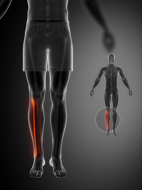
Human skeleton anatomy Tibia Bone 3D Rendering For Medical Concept

Hip, leg, thigh and pelvis injury medical x-ray test scan image of old age senior adult.
X-ray scan of human chest and lungs.

Hip x-ray of human
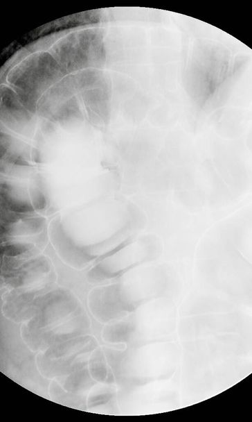
X-ray examination Spot film barium enema examination (lower gastrointestinal).

Articular capsule Anatomy For Medical Concept 3D Illustration

Hip joint replacement xray showing ball and socket joint's titanium screw implant in medical orthpodedics scan.

Human Skeleton Vertebral Column Cervical Vertebrae Anatomy 3D Illustration

Hamstring muscle group anatomy model. 3d illustration.

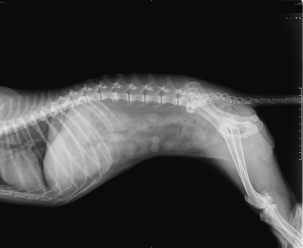
Thorax lateral x-ray film of cat

Medical hospital x-ray hips spine pelvis MRI traumatology scan.
Spine and pelvis of a human body on x-ray

3d rendered illustration - femur bone - front view

Human skeleton anatomy Humerus Bone 3D Rendering For Medical Concept

Human skeleton: pelvis and sacrum. Medically accurate 3D illustration

Male bones anatomy pelvis
Hip replacement xray orthopedic medical x-ray Traumatology test scan image of old age senior adult.

The spine of a human body on x-ray

Hip joint, spine and pelvis xray scan test results for adult patient.

Hip joint replacement xray showing ball and socket joint's titanium screw implant in medical orthpodedics scan.
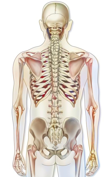
Bone system: human skeleton in posterior view.