


Filters

Stock royalty-free photos and images of Protist
Discover unlimited high resolution images of Protist and stock visuals for commercial use.

Foraminifera are microscopic marine amoeboid protists with calcareous tests. Genera Calcarina, Sorites, and Baculogypsina from left to right. The width of the view is 10 mm.

Microscopic view of Noctiluca scintillans, commonly known as the sea sparkle.

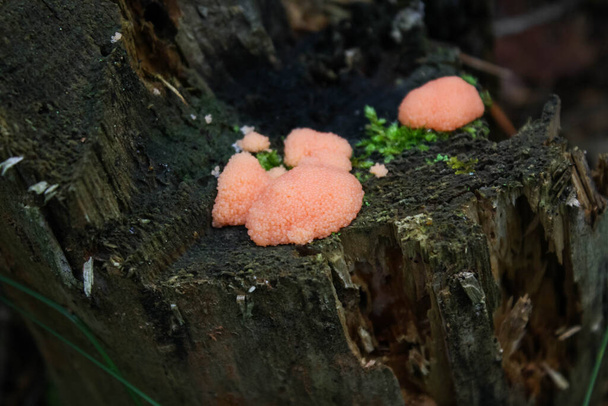
True Slime Mold of the genus Fuligo

Leucarpus fragilis Eggshell Insect-egg or Fragile Yellow Slime Mold is a Protist kingdom organism a very common myxomycete in the bush when the environment is humid and temperate natural light

Cribraria argillacea Lead shot slime mold myxomycete even young gray and looks like lead balls growing on a pine trunk natural light

Retinal scar in toxoplasmosis, a disease caused by the single-celled protozoan Toxoplasma gondii, scientific illustration, fluorescein angiography

Retinal scar in toxoplasmosis, a disease caused by the single-celled protozoan Toxoplasma gondii, ophthalmoscope view, 3D illustration

3d illustration of flower vase decoration isolated on black background

Protozoology typography, wordcloud, wordart,

Microscopic Life. Microscope. Single Cell Animal. Living Organism. Seen through a microscope. Animal Life. Microscopic Rotifer or Rotifera under a microscope, freshwater bentic organism filtering water. Microscopic Life seen through a Microscope.

Plasmodium of a slime mold (Badhamia utricularis) spreading in a wave across a piece of rotting wood
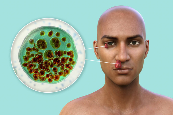
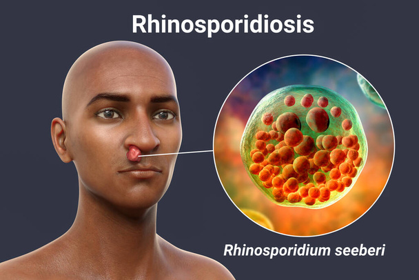
Nasal and ocular rhinosporidiosis in a patient, 3D illustration. A disease caused by Rhinosporidium seeberi parasite, leads to formation of polypoidal masses in nasopharynx and conjunctiva
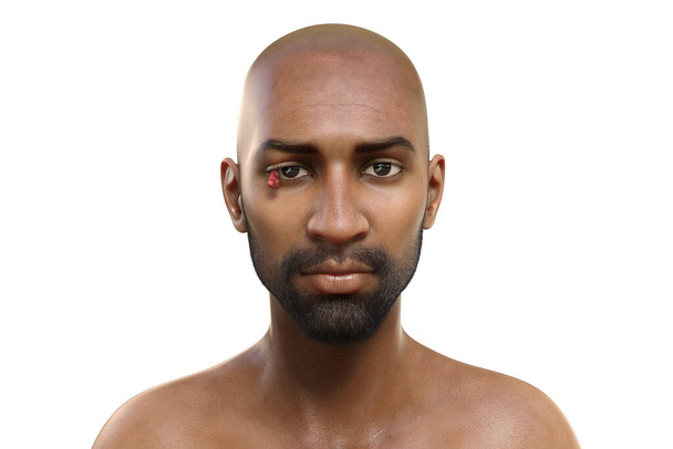
Ocular rhinosporidiosis in a patient, 3D illustration. A disease caused by Rhinosporidium seeberi parasite and leads to formation of polypoidal masses in nasopharynx and conjunctiva

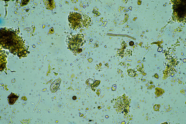
Arcella, fungi and nematode in a soil sample on a farm in australia

Host cells with spores (mold) are inside wood under the microscope for education.

Host cells with spores (mold) are inside wood under the microscope for education.

Host cells with spores (mold) are inside wood under the microscope for education.

Host cells with spores (mold) are inside wood under the microscope for education.

Living Vorticella is a genus of protozoan under microscop view.

Microscopic Life. Microscope. Single Cell Animal. Living Organism. Seen through a microscope. Animal Life. Microscopic Rotifer or Rotifera under a microscope, freshwater bentic organism filtering water. Microscopic Life seen through a Microscope.

Ossicaulis lachnopus is a reddish-white mushroom that grows on decaying tree trunks such as poplars in autumn when the humidity and temperature are mild light by flash

Host cells with spores (mold) are inside wood under the microscope for education.

Host cells with spores (mold) are inside wood under the microscope for education.

Protozoa and Green Algae in waste water under the microscope.

Vorticella is a genus of protozoan under microscop view.

Sporangia of the Many Headed Slime of the species Physarum polycephalum scattered on dry leaves on the ground

Rhinosporidium seeberi parasite, the causative agent of rhinosporidiosis, disease with formation of polypoidal masses in nasopharynx, 3D illustration shows mature (big) and immature (small) sporangia

Arcella, fungi and nematode in a soil sample on a farm in australia
Arcella, fungi and nematode in a soil sample on a farm in australia

Arcella, fungi and nematode in a soil sample on a farm in australia

Wolfs milk slime growing on a tree stump

Microscopic photograph of a CILIATE a Protozan Phylum Ciliophora. Photographed through a Microscope at 1000 times its actual size. Microscopic life. Living Organism.

Close-up image of the yellow fruiting body of a golden apple slime mold (Arcyria pomiformis) emerging from a piece of rotten wood. This specimen was about 1 millimeter tall

Protozoa and Green Algae in waste water under the microscope.

Host cells with spores (mold) are inside wood under the microscope for education.

Host cells with spores (mold) are inside wood under the microscope for education.

Host cells with spores (mold) are inside wood under the microscope for education.

Desmids are a common group of freshwater single-celled algae that have intricate cell walls
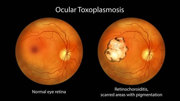
Retinal scar in toxoplasmosis, a disease caused by the single-celled protozoan Toxoplasma gondii, and the same healthy eye retina for comparison, ophthalmoscope view, scientific illustration
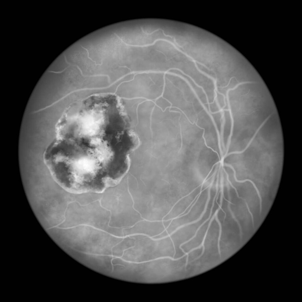
Retinal scar in toxoplasmosis, a disease caused by the single-celled protozoan Toxoplasma gondii, scientific illustration, fluorescein angiography
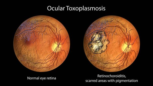
Retinal scar in toxoplasmosis, a disease caused by the single-celled protozoan Toxoplasma gondii, and the same healthy eye retina for comparison, ophthalmoscope view, 3D illustration

Arcella in a tissue sample in a lab

Slime mold plasmodium (Badhamia utricularis) networked to feed on rolled oats and a piece of mushroom

Arcella, fungi and nematode in a soil sample on a farm in australia

Arcella, fungi and nematode in a soil sample on a farm in australia
Nasal rhinosporidiosis in a patient, 3D illustration. A disease caused by Rhinosporidium seeberi parasite, leads to formation of polypoidal masses in nasopharynx and conjunctiva

Nasal rhinosporidiosis in a patient, 3D illustration. A disease caused by Rhinosporidium seeberi parasite, leads to formation of polypoidal masses in nasopharynx and conjunctiva

Retinal scar in toxoplasmosis, a disease caused by the single-celled protozoan Toxoplasma gondii, scientific illustration, fluorescein angiography

Arcella, fungi and nematode in a soil sample on a farm in australia

Arcella, fungi and nematode in a soil sample on a farm in australia

Arcella, fungi and nematode in a soil sample on a farm in australia
Pinc Slime Tubulifera arachnoidea, formerly known as Tubifera ferruginosa. Macro

Microscopic Life. Microscope. Single Cell Animal. Living Organism. Seen through a microscope. Animal Life. Microscopic Rotifer or Rotifera under a microscope, freshwater bentic organism filtering water. Microscopic Life seen through a Microscope.

Microscopic Life. Microscope. Single Cell Animal. Living Organism. Seen through a microscope. Animal Life. Microscopic Rotifer or Rotifera under a microscope, freshwater bentic organism filtering water. Microscopic Life seen through a Microscope.

Arcella, fungi and nematode in a soil sample on a farm in australia

Host cells with spores (mold) are inside wood under the microscope for education.

Host cells with spores (mold) are inside wood under the microscope for education.

Vorticella is a genus of protozoan under microscop view.

Desmids are a common group of freshwater single-celled algae that have intricate cell walls

Esmids are a common group of freshwater single-celled algae that have intricate cell walls

Sporangia of the Many Headed Slime of the species Physarum polycephalum scattered on dry leaves on the ground

Ocular rhinosporidiosis in a patient and the same healthy man, 3D illustration. A disease caused by Rhinosporidium seeberi parasite with formation of polypoidal masses in nasopharynx and conjunctiva