


Filters

Stock royalty-free photos and images of Eixo direito
Discover unlimited high resolution images of Eixo direito and stock visuals for commercial use.

Right angle, construction, ruler on white background

Macro shot of a red flamingo flower (lat. Anthurium) (Selective Focus, Focus on the spadix from the middle until the right upper corner of picture)

Four anatomical malformations of tetralogy of Fallot: 1 aortic straddling; 2 ventricular septal defect; 3 right ventricular hypertrophy and 4 pulmonary artery stenosis.

Right axis deviation can be seen both in right ventricular hypertrophy and in some healthy people, who have no ECG abnormalities except for right axis deviation.

When the area or mass of blood supplied to the posterior LV by the RCA exceeds that of the RV, acute inferior MI also occurs with a lead II elevation amplitude greater than that of lead III.

When the frequency of sinus rhythm is less than 60 beats per minute, it is called sinus bradycardia. Severe sinus bradycardia should be suspicious of sick sinus syndrome.

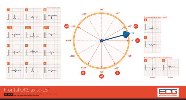
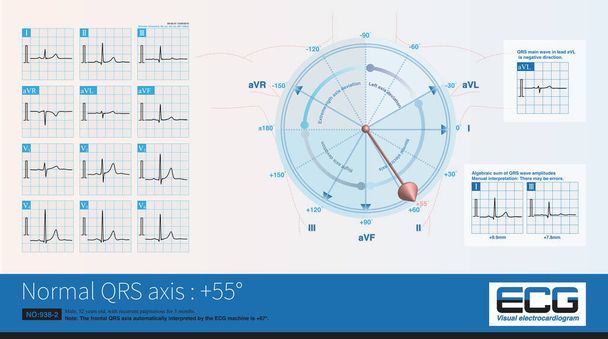
When the frontal QRS axis is located at +76, it is located on the negative side of the aVL lead axis, and the QRS main wave in lead aVL is negative.The highest amplitude of the QRS wave in the limb lead is the aVF lead.
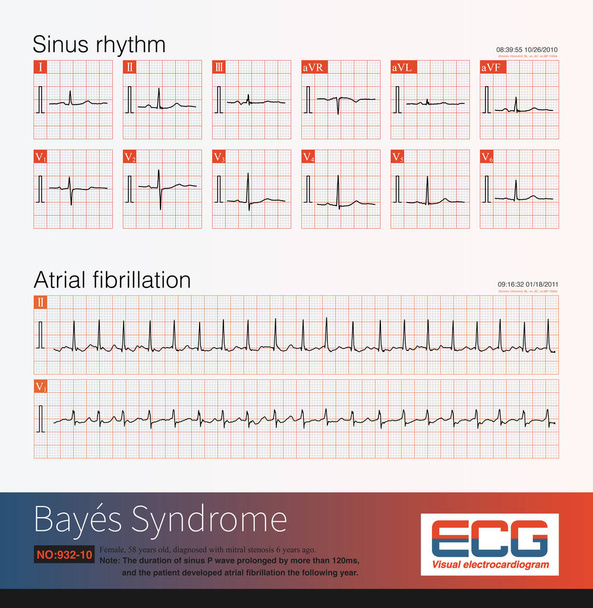
A pre elderly woman suffered from mitral stenosis. In 2010, her electrocardiogram showed sinus rhythm and left atrial abnormality, and in 2011, she progressed to atrial fibrillation.
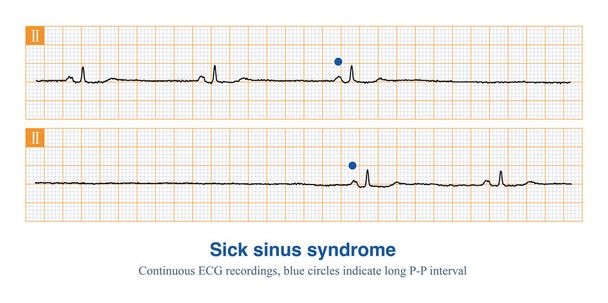
When two or more types of slowing sinus arrhythmias, such as sinus bradycardia, sinoatrial block, and sinus arrest, occur simultaneously, one should be alert to sick sinus syndrome.
Female, 27 years old, clinically diagnosed as uremia for 5 years, received dialysis treatment.Chest lead QRS wave showed low voltage.
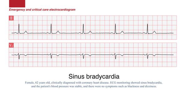
Sinus heart rate less than 60 beats per minute in adults is called sinus bradycardia. A sinus heart rate of 40 to 50 beats per minute is moderate sinus bradycardia, and patients may have symptoms such as palpitations and dizziness.

On a 12 lead electrocardiogram, analyzing the morphology and polarity of ectopic P-waves can determine the approximate anatomical location of ectopic atrial foci.

Champion South Texas axis buck standing looking to the right
Sometimes, the ST segment of the electrocardiogram in young and middle-aged women shows an upward oblique depression, many of which belong to physiological ST segment depression.
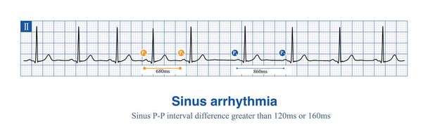
Sinus arrhythmia is a very common benign arrhythmia that does not require treatment. It can be diagnosed with a sinus cycle difference of more than 120ms or 160ms.
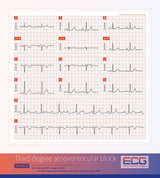
Third degree atrioventricular block in young women may be congenital, with the block located on the atrioventricular node or above bifurcation of the His bundle.

Moving through neon straight tunnel made of simple square green gates and curved narrow yellow lines flowing on black background. Colorful magnetic rhombus tunnel.

Male, 39 years old, paroxysmal palpitations for 5 years. ECG showed B type ventricular preexcitation.This diagram shows how to measure ventricular preexcitation components simultaneously.

The frequency of sinus impulses exceeds 100 beats per minute, and the electrocardiogram diagnosis is sinus tachycardia. The important differential diagnosis is atrial tachycardia.

Long term mitral stenosis leads to pulmonary hypertension, which eventually leads to increased right ventricular afterload and right ventricular hypertrophy.ECG showed right axis deviation.
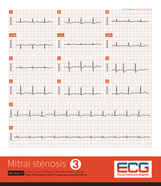
Female, 53 years old, diagnosed with mitral stenosis 2 years ago. When this ECG was taken, the patient still maintained sinus rhythm.Note that the P wave duration was widened.

Female, 52 years old, diagnosed with mitral stenosis 1 years ago. When this ECG was taken, the patient still maintained sinus rhythm.Note that the P wave duration was widened.
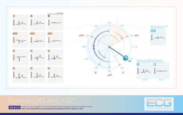
When the frontal axis is + 32 , the maximum QRS depolarization potential is closest to the axis of lead II, so the R-wave amplitude of lead II is the highest.

When ectopic impulses from the anterior wall of the right atrium produce a completely negative P wave in lead V1, the posterior wall ectopic impulse produces a positive and negative biphasic P wave.

SQT sign is an electrocardiogram change of right heart disease, which is not only seen in acute pulmonary embolism, but also in other diseases affecting the right heart.

Respiratory sinus arrhythmia is diagnosed when the heart rate increases during inspiration and slows down during expiration, and the difference between the P-P interval exceeds 120 or 160 ms.

When the electrical axis of the electrocardiogram is severely left deviation, especially near the negative side of the aVF lead axis, significant S-waves appear in leads I, II, and III.

When the frontal QRS axis is at +62, the QRS amplitude of lead is the highest.The frontal electrical axis is almost perpendicular to aVL lead, so the algebraic sum of QRS wave amplitude in aVL lead is almost zero.
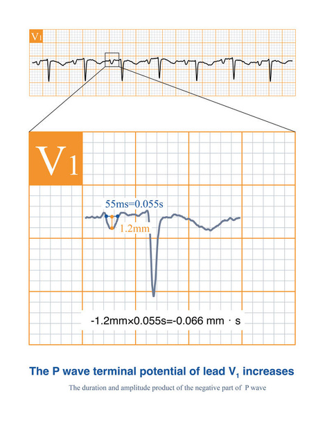
When the left atrium is abnormal, the absolute value of the duration and amplitude product of the negative part of lead V1 P-wave will increase, indicating dysfunction of the left atrium.