


Filters

Stock royalty-free photos and images of Biopsja
Discover unlimited high resolution images of Biopsja and stock visuals for commercial use.
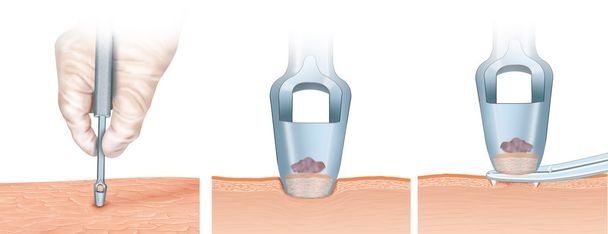
Illustrations on surgical intervention punch
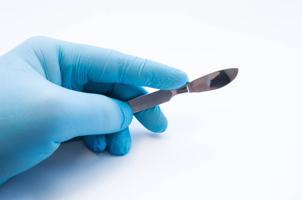
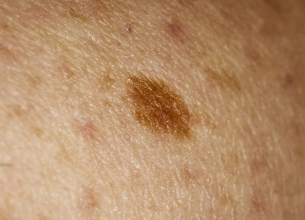
Hand holding scalpel. Palm of surgeon dressed in blue glove holding scalpel. Concept photo for surgeries, procedures, treatment, plastic surgery operation, work of surgical departments and hospitals
Biopsy medical concept image with icons and doctors on background

All products lactose free

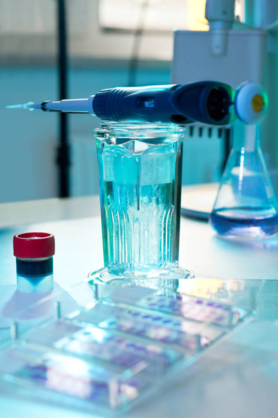
Close up scientist woman research carrying chemistry liquid with microscope equipment for research experiments to test tube at science laboratory
Histochemical staining of tissue samples

Human anatomy exploded view, 3D illustration

Dermatologist takes sample for analysis from under the patient's toenail. Dermatology, fungal infection. Close up

Science concept. Laboratory equipment composition. Test tubes on blue background.

Cervical Cancer Word Cloud Concept in red caps with great terms such as prevention, women, virus and more.

Medical Concept: Black Chalkboard with Melanoma. Medical Concept: Melanoma - Text on Black Chalkboard with Red Stethoscope. 3D Rendering.

Melanoma, word cloud concept on white background.

Cells in reproductive female cytology and histology concept medical scinece.

Liver cancer hepatoma with t-cell realistic colseup view 3d illustration
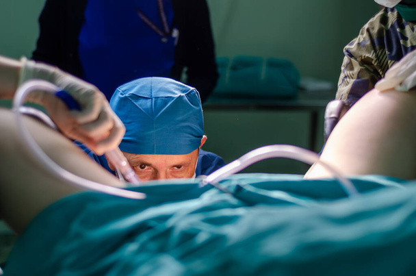
Surgeon doctor in operating room during gynecological biopsy

Bone marrow core biopsy pathology - disseminated Histoplasmosis

Prostate hyperplasia. Photomicrograph showing dilated glands, enfolding of the glandular epithelial cells forming papillary projections into the gland lumen, cystic dilatation

Skin cancer cells isolated on white background, 3D illustration

Checking benign moles. Close up detail of the bare skin on a man back with scattered moles and freckles. Sun effect on skin. Pigmentation. Birthmarks on skin

Selective focus of blood collection during blood donation and ball in palm for squeezing

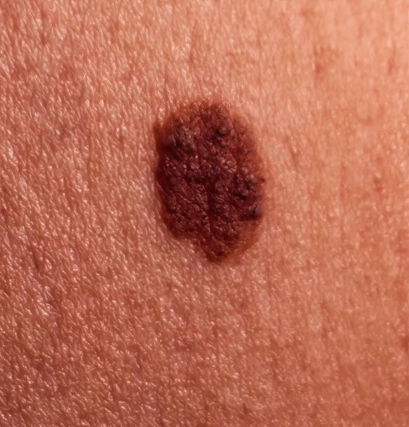
Mole birthmark nevus macro photo on human skin. Close up

Rotary Microtome Section for diagnosis in pathology make microscope slide histology. Human tissue equipment

Squamous epithelial cells under microscope view for education histology. Histological for human physiology.

Medical Scissors over the white background.

Rotary Microtome Section for diagnosis in pathology make microscope slide histology. Human tissue equipment

Science concept. Laboratory equipment composition. Test tubes on blue background.
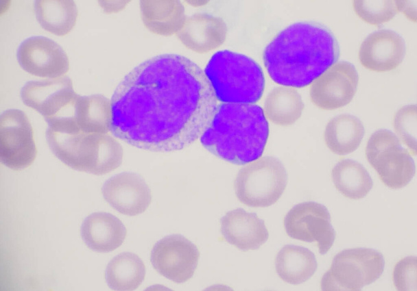
Group of blast cells in leukemia blood smear background.

Papilloma on human skin - benign tumor in the form of mole, nevus Papillomatosis medicine

Colorectal cancer, intestinal carcinoma, bowel neoplasia, 3D illustration showing malignant tumor in intestine

Melanoma cell a type of skin cancer closeup view 3d illustration

"The Human Digestive System. 3D illustration of the colonoscopy Procedure"

Anatomy and Histological Bone, Elastic cartilage human and Joint of human foetus under the microscope for education.

Photomicrograph of colon adenocarcinoma, illustrating malignant glandular cells characteristic of colon cancer.
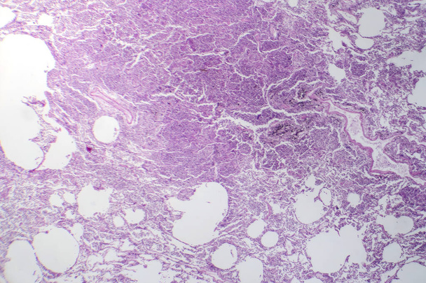
Photomicrograph of interstitial pneumonia, showing inflammation and fibrosis in the lung's interstitial tissue.

Photomicrograph of nasal polyps, displaying abnormal tissue growth in the nasal passages often causing congestion and discomfort.

Colorectal cancer, malignant tumor in intestine, Endoscope inside colonoscopy, gut intestine, Colon polyp removal, colonic polyps search, Polypectomy, intestinal carcinoma, bowel neoplasia, 3d render
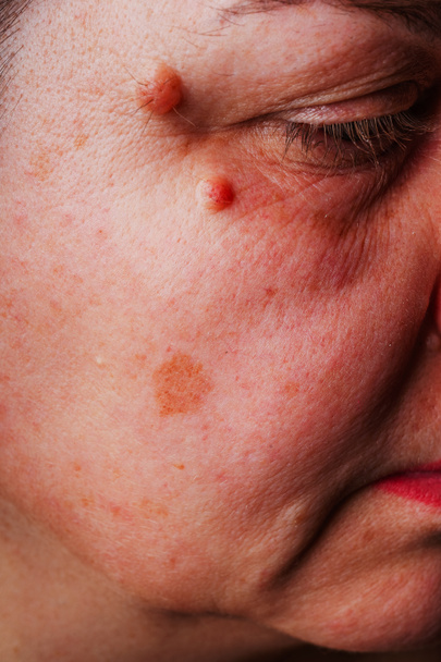
Large moles near the eye close-up. Red large round moles on the woman's face. Inflammation of the tear duct. A woman's reddened eye. Spots on the face and under the eyes in liver diseases

Abnormal cells in pleural fluid wright staining.

Young woman doctor's hands close up preparing for an ultrasound device scan.
Nuclear medicine medical concept image with icons and doctors on background
Heart surgery medical concept image with icons and doctors on background
Anesthesiology medical concept image with icons and doctors on background

Laboratory microscope for blood analysis
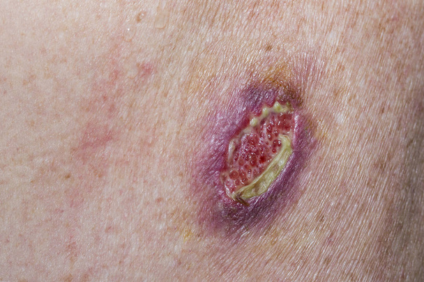
Wound dehiscence - open wound from unsuccessful medical stitches in a horizontal format

Men in a laboratory microscope with microscope slide in hand.

Cancer cell growth in culture,Electron Microscope

Close up microscope equipment for research experiments in science laboratory

Young woman pointing at dark brown moles on her back for self-exams skin. Being aware of changes in your moles to detecting skin cancer, especially malignant melanoma. Skin disease concept.

Mature dendritic cell - super closeup view 3d illustration
Large mole on human skin. Mole removal concept. Close-up, selective focus

Acute lymphoblastic leukemia (ALL) cancer cells in the blood flow - closeup view 3d illustration

Lipoma, a growth of fat tissue under human skin, cross-section view, 3D illustration

Cancer cell in the moment that divides, 3D Illustration
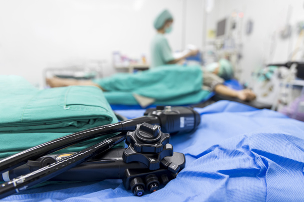
Medical colonoscopy or gastroscopy instrument on surgical table inside operating room in hospital.Endoscopic technology for cancer screening with blur background and space.Medical device for surgeon.
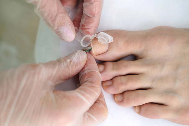
Dermatologist takes sample for analysis from under the patient's toenail. Dermatology, fungal infection. Close up
Large mole on human skin. Mole removal concept. Macro photo, selective focus

Close up detail of the bare skin on a man back with scattered moles and freckles. Checking benign moles. Sun effect on skin. Birthmarks on skin

Crimson red spirochetes is a spiral shaped bacterium that is Gram negative and generally undergoes anaerobic phototrophic living.Magnify 1000x

Cells of a human thyroid gland with goiter caused by deficiency of iodine under a microscope.

Areolar connective tissue under the microscope view. Histological for human physiology.

Surgical instrument on a white background

Surgical instrument on a white background

Human anatomy exploded view, 3D illustration

Medical hospital mammography x-ray breast cancer mammogram scan.

Writing note showing Melanoma. Business photo showcasing A malignant tumor associated with skin cancer Benign moles.

Scientist hand with dropping chemical liquid with scientist carrying research chemistry liquid to test tube with microscope equipment for research experiments in science laboratory research and experiment concept

Text sign showing Lymphoma. Conceptual photo Cancer that begins in infection fighting cells of the immune system.

Word writing text Melanoma. Business concept for A malignant tumor associated with skin cancer Benign moles.

Conceptual hand writing showing Lymphoma. Business photo text Cancer that begins in infection fighting cells of the immune system.

Melanoma - Printed Diagnosis with Blurred Text. Melanoma, Medical Concept with Pills, Injections and Syringe. 3D. Toned Image

Tumor - Printed Diagnosis with Blue Pills, Injections and Syringe. Medical Concept with Selective Focus. 3D Render.

Tumor - Printed Diagnosis with Blurred Text on Red Background with Specs. Medicine Concept. Tumor - Medical Concept on Red Background with Blurred Text and Composition of Eyeglasses. 3D Rendering.

Colon Cancer word cloud hand writing concept on white background.

Prognosis, word cloud concept on white background
Skin Tags, word cloud concept on white background.
Skin Cancer, word cloud concept on black background.

Cervical Cancer Word Cloud Concept on a 3D cube Blackboard with great terms such as prevention, women, virus and more.

Stitch Scars on the Face and bald head of young man

Handwriting Concept - Color Image

Mammography breast screening device in hospital laboratory. Health care, medical technology, hi-tech equipment concept.
Aesthetic surgery text medical concept image with icons and doctors on background
Neurology medical concept image with icons and doctors on background

Asian/Indian male scientist or doctor or science student experimenting with microscope and chemicals, laptop and smartphone in a lab

Science concept. Laboratory equipment composition. Test tubes on blue background.

Cross section through cells of a human thymus gland under the microscope.

Laboratory equipment composition. Science concept.

Text sign showing Cervical Cancer. Business photo showcasing type of cancer that occurs in the cells of the cervix

Human lung tissue with dust and coal particles (miner lung) under the microscope.

Human lung tissue with dust and coal particles (miner lung) under the microscope.

Handsome male doctor discussing x-ray of a foot with his colleague. Young practitioner examining x-ray scan of a patient, talking to his co-worker. Medicine, technology concept
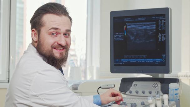
Male doctor examining results of ultrasound scanning of a patient. Bearded male practitioner using transducer for ultrasound examination. Cheerful doctor smiling to the camera

A section of a mouse kidney under the microscope.

Dermatologist examining patient with magnifying glass in clinic, closeup view

Cytology typography, wordart, wordcloud, cytology, medical, science, pathology

Patrology typography, wordcloud, wordart, pathology, medical, medicine, science

Woman's oily skin with acne problems. Scars and wounds on the face. Health care photo. Medical balm for pimple.

Conceptual display Cervical Cancer, Word Written on type of cancer that occurs in the cells of the cervix Abstract Sending Multiple Messages Online, Typing Group Lessons

Conceptual display Cervical Cancer, Business approach type of cancer that occurs in the cells of the cervix Hands Pointing Pressing Computer Keyboard Keys Typewriting New Ideas.

Big birthmark on Asian boy stomach. Close-up of brown birthmark on Asian fat boy.

Liquid based cytology microscope slide for pap smear test. Cervical cancer concept. Medical concept.