


Filters

Stock royalty-free photos and images of Retina
Discover unlimited high resolution images of Retina and stock visuals for commercial use.
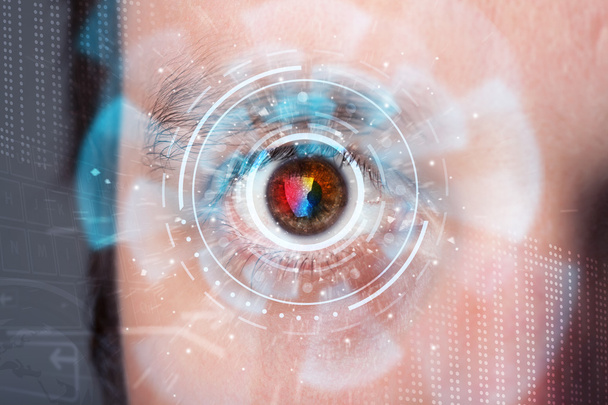
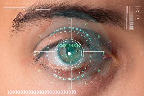
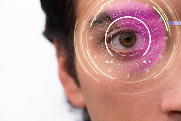
Futuristic modern cyber man with technology screen eye panel concept

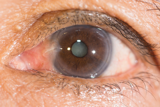
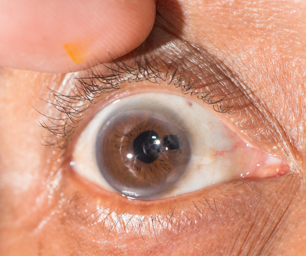
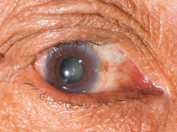
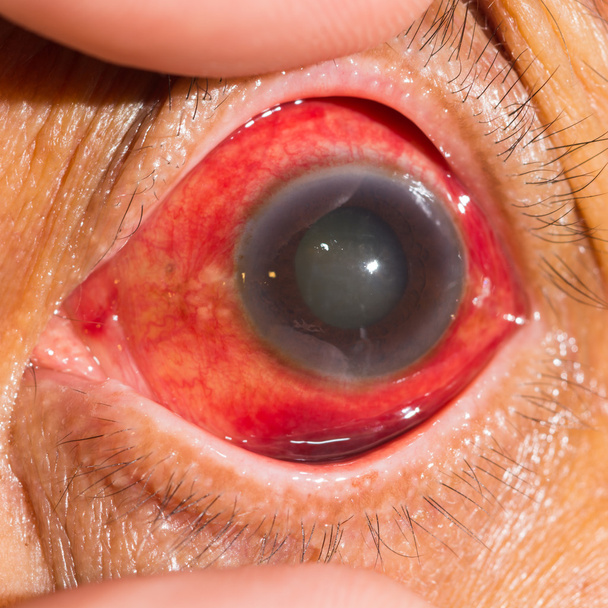
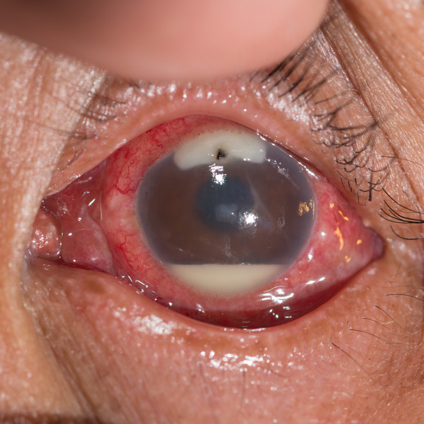
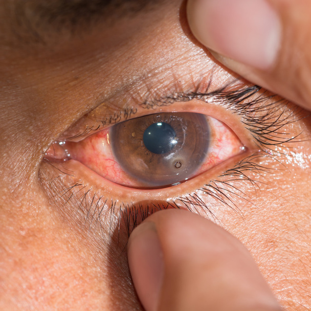
Close-up of the neovascular glaucoma.During eye examination

Futuristic modern cyber man with technology screen eye panel concept
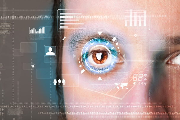
Futuristic modern cyber man with technology screen eye panel concept

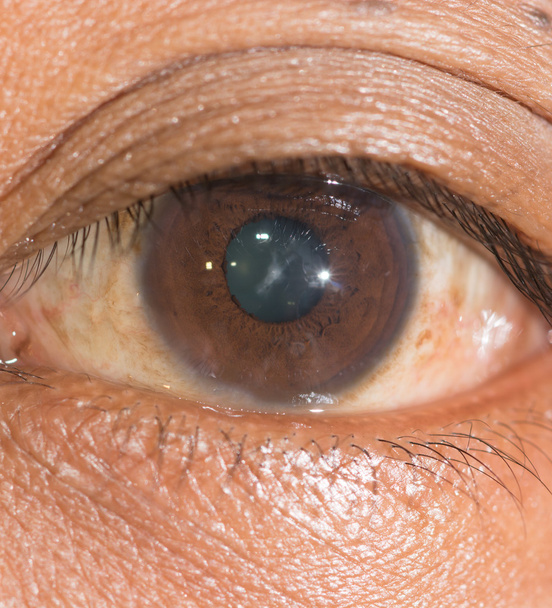
Close up of the senile during eye examination.

Close up of the trichiasis during eye examination.

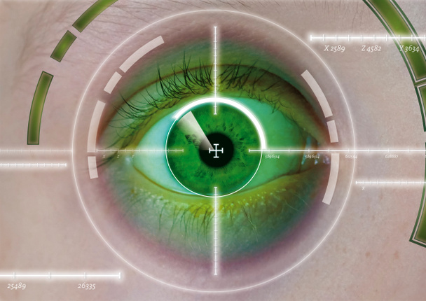
Future woman with cyber technology eye panel concept

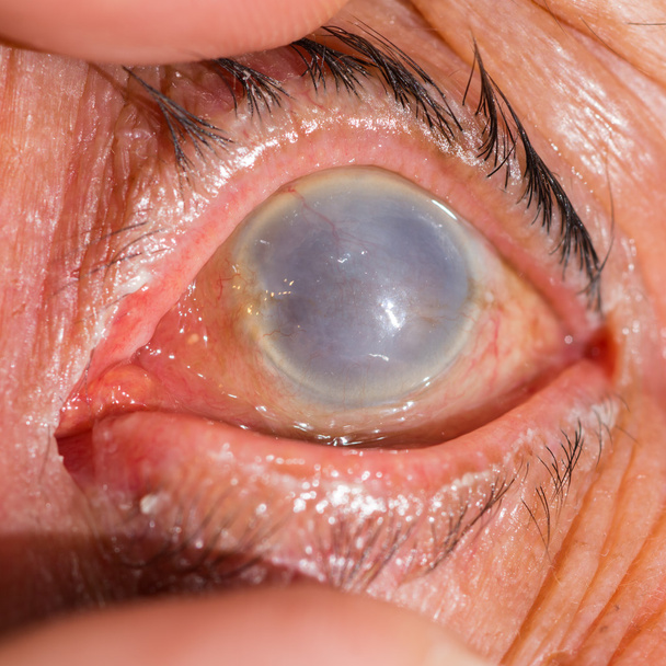
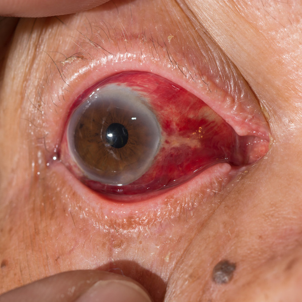
Close up of the phthisis bulbi during eye examination.

Close up of the sub conjunctival heamorrhage during eye examination.

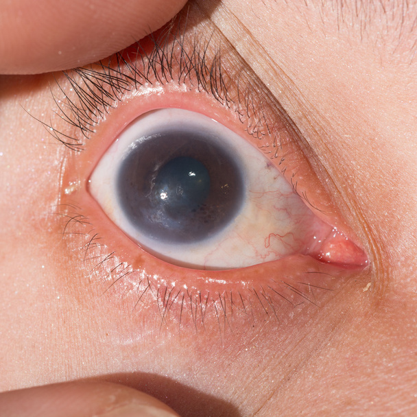
Close-up of the acute glaucoma.During eye examination.

Close up of the retained metallic corneal foreign body during eye examination.

Close up of the severe bacterial corneal ulcer during eye examination.

Eye anatomy diagram,3 d rendering image .
Close up of the pterygium and senile cataract during eye examination.

Close up of the senile during eye examination.
Futuristic modern cyber man with technology screen eye panel concept
Close up of the repaired cornel penetrating wound during eye examination.
Close up of the anterior sutural cataract during eye examination.

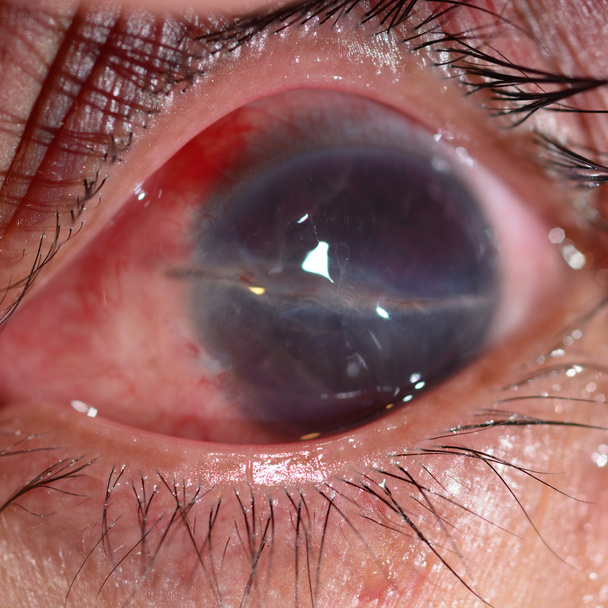
Close up of the acute angle closure glaucoma during eye examination.

Close up of the cataract during eye examination.

Close up of the dilated pupil eye during eye examination.

Close up of the mature cataract during eye examination.
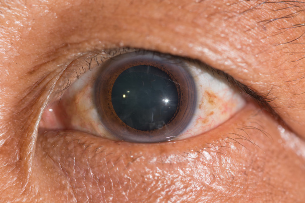
Close up of the dilated pupil during eye examination.

Close up of eye examination, Herpes keratitis.
Concept of sensor implanted into human eye
Close up of the trichiasis during eye examination.

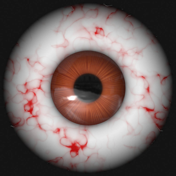
Gouged bloodshot eyeball on white background

The close up shot of human eye. The human eye is a paired sense organ that reacts to light and allows vision.

Close up of the senile cataract during eye examination.
Eye laser surgery, Diopter, eye, laser, correction,

Close up of the post Penetrating Keratoplasty during eye examination.
Close up of the keratitis during eye examination.

Close up of the pseudo phakic bullous keratopathy during eye examination.

Asian women being futuristic vision, digital technology screen over the eye vision background, security and command in the accesses. surveillance and sefety concept

Close up of acute viral conjunctivitis during eye examination.
Close up of the total opacity cornea during eye examination.

Close up of the corneal scar during eye examination.
Eyeball with iris pupil

Close up of the advance pterygium during eye examination.
Close-up of the acute glaucoma.During eye examination.

Close up of acute anterior uveitis during eye examination.

A texture of a blue eye with lots of detail.
Close up of the traumatic corneal laceratiion during eye examination.

Iris eyes. Human iris with blood veins. Eye illustration. Creative digital graphic design

Human Eye Dissection Anatomy

Concept of sensor implanted into human eye

Close up of the pterygium during eye examination.

Close up of acute anterior uveitis during eye examination.
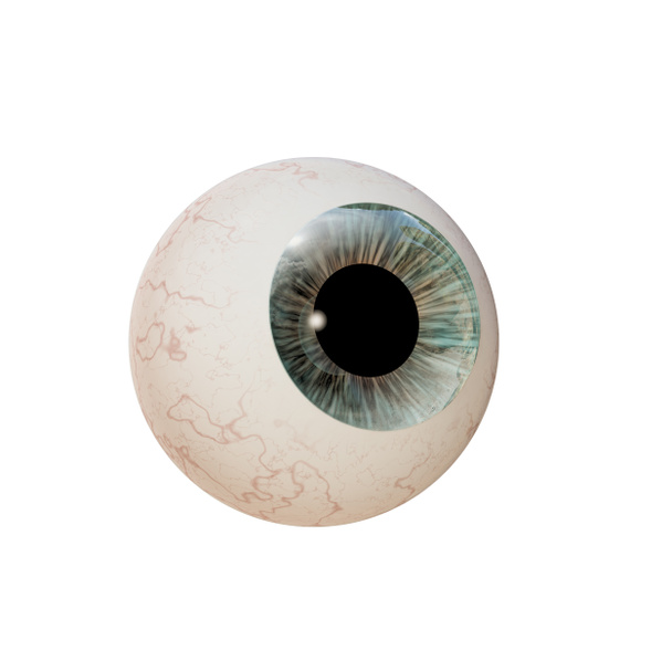
Eyeball isolated on white background
Close up of the posterior synechiae during eye examination
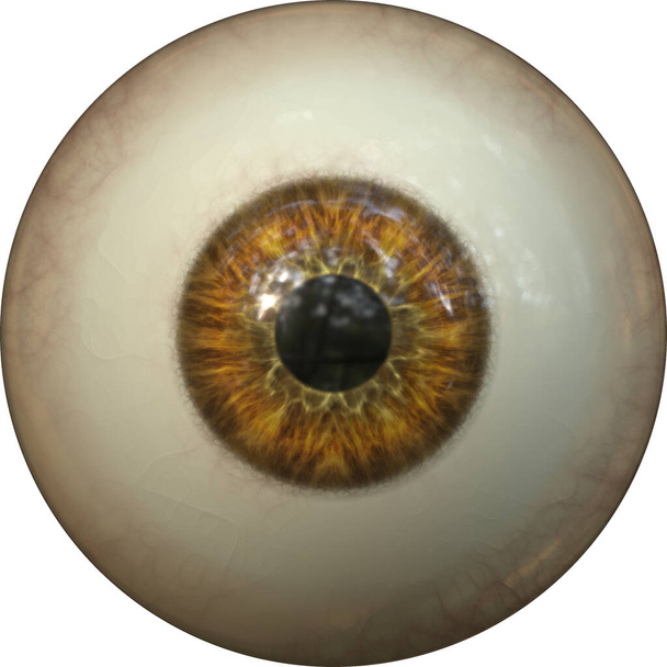
Eyeball with iris pupil

Illustration of human eyeball with green iris

Eye: detachment of the retina, which detaches from the underlying choroid.

Future woman with cyber technology eye panel concept
Concept of sensor implanted into human eye

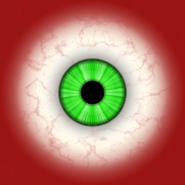
Isolated eye. Raptor purple eye with large pupil and bright red retina in background. Dark iris around pupil.
Close up of the retained metallic foriegn body with hypopyon during eye examination.
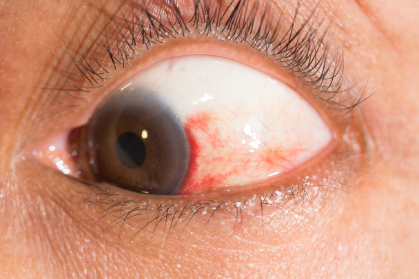
Close up of the subconjunctival hemorrhage during eye examination.

Isolated eye. Raptor purple eye with large pupil and bright red retina in background. Dark iris around pupil.

Close up of the corneal ulcer during eye examination.
Close up of the subconjunctival hemorrhage during eye examination.

Biometrics, eye scanning and recognition concept.
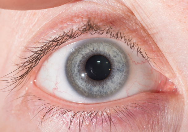
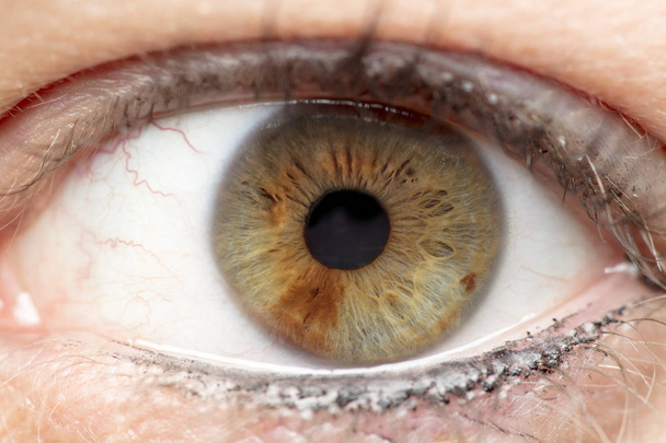
Close up of normal grey iris during eye examination.

Proliferative diabetic retinopathy, illustration showing neovascularization (formation of new vessels) in the optic disk. Fundoscopic examination of the eye retina in diabetes mellitus
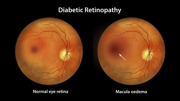
Diabetic macular edema (DME), illustration showing normal eye retina and retina with macula edema. Fundoscopic examination of the eye retina in diabetes mellitus

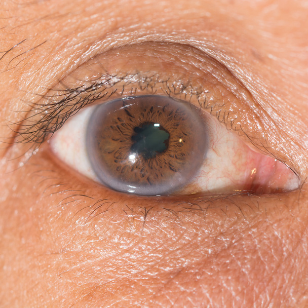
Senile cataract during eye examination close up
Eyeball isolated on red background

Eyeball closeup

Close up of the periorbital ecchymosis during eye examination.

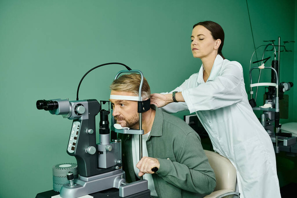
Human eyes retina, professional ophthalmological equipment. Concept: Ophthalmology Field of medicine

Closeup view of eyeball

Illustration of human eyeball with blue iris

The concept of sensor implanted into human eye

Sagittal view of the eye anatomy showing lens, retina, cornea, iris, choroid. .

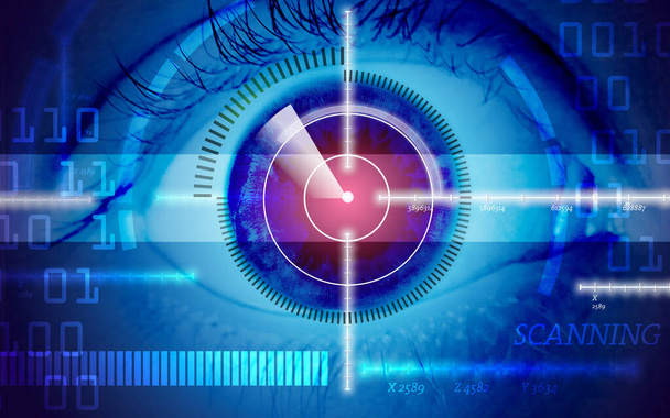
Biometric scan of the female eye close-up. The concept of modern virtual reality
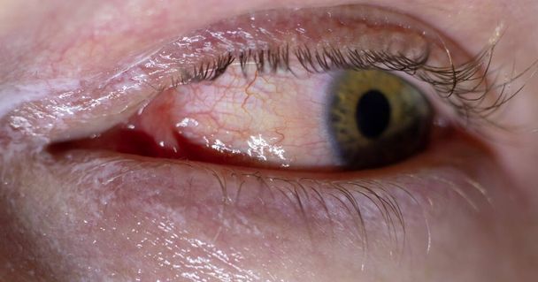
Inflamated eye infection. Red bloody eye closeup macro result of allergies and tiredness. Watery red skin around with healing creme applied

Close up of the subconjunctival hemorrhage during eye examination.

Close up of the cortical cataract during eye examination.

Close up of the periorbital ecchymosis during eye examination.
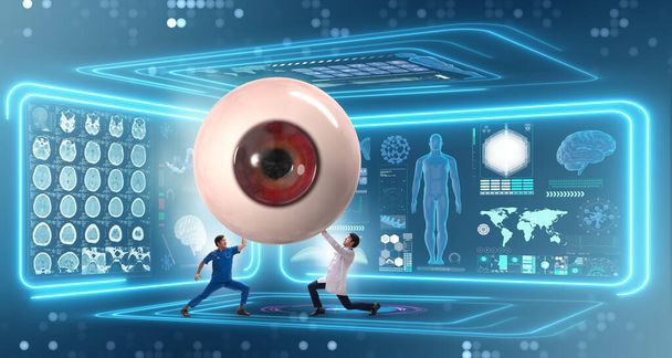
Doctor examining giant eye in medical concept

Smooth orange eye. Animal 3D eye with large pupil and dark retina in background.

Iris eyes. Human iris with blood veins. Eye illustration. Blue eye. Creative digital graphic design

Close up of acute anterior uveitis during eye examination.
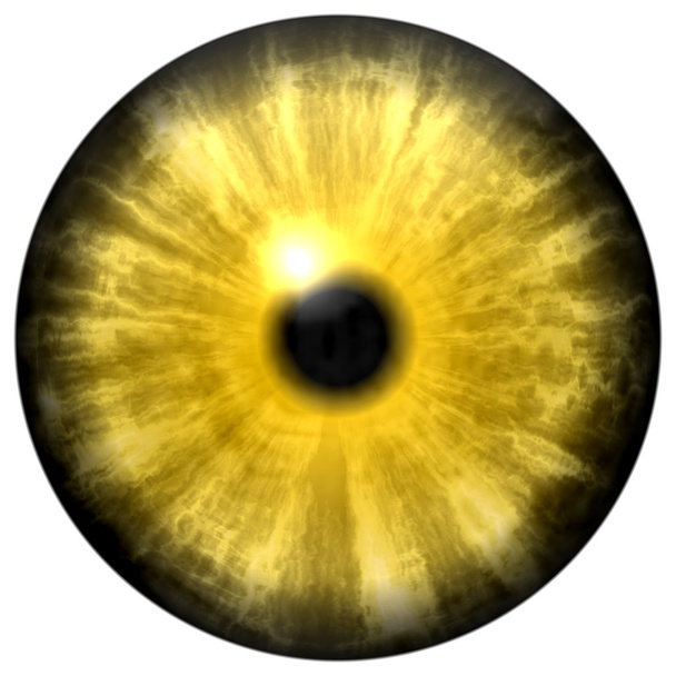
Yellow animal eye with small pupil and black retina in background. Dark colorful iris around pupil, detail view into eye bulb.

Close-up of the neovascular glaucoma.During eye examination

The eye illustration in medical concept - 3d rendering

Close up of episcleritis during eye examination.
Eye scanner - internet security

Non-proliferative diabetic retinopathy, illustration showing normal eye retina and retina with hard exudates, microaneurysms, dot haemorrhages, flame-shaped and splinter retinal haemorrhages
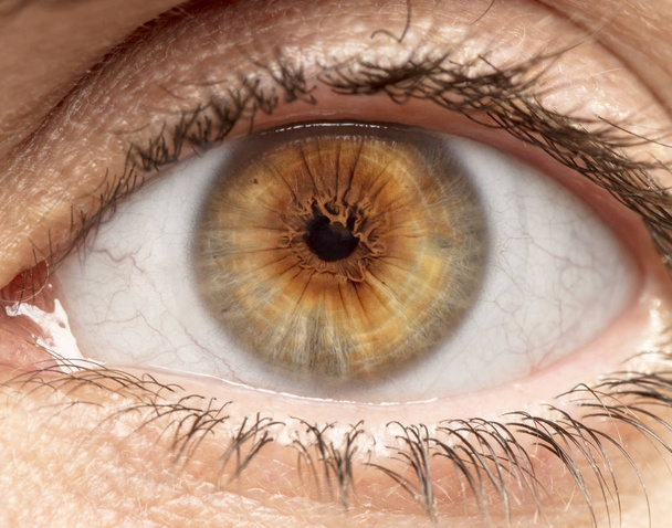
Macro photo of human eye, iris, pupil, eye lashes, eye lids.
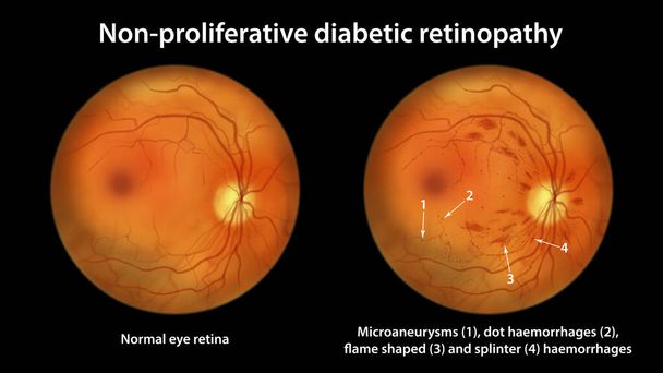
Non-proliferative diabetic retinopathy, illustration showing normal eye retina and retina with microaneurysms, dot haemorrhages, flame-shaped and splinter retinal haemorrhages, ophthalmoscope view
Close up of the metallic foreign body on cornea "rust ring" during eye examination.

Close up of the congenital corneal problem during eye examination.
Macro photo of human eye, iris, pupil, eye lashes, eye lids.

3D illustration - Human eye isolated on black background

A human eye recognition image with data stream design in black and white

3D illustration - Blue human eye isolated on white background

Close up of sub conjunctival hemorrhage during eye examination.
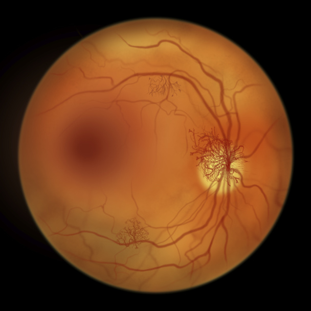
Proliferative diabetic retinopathy, illustration showing neovascularization in the disk and other sites, and macula edema. Fundoscopic examination of the eye retina in diabetes mellitus