


Filters

Stock royalty-free photos and images of Слайды микроскопа
Discover unlimited high resolution images of Слайды микроскопа and stock visuals for commercial use.

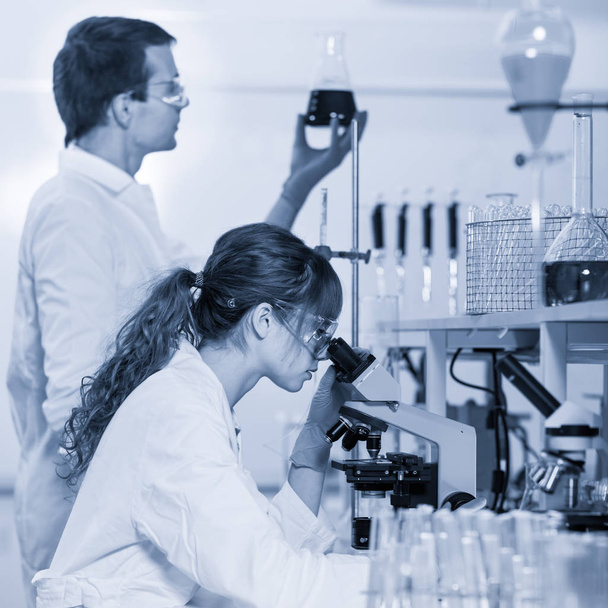
Life scientist researching in laboratory. Attractive young male scientist looking at the microscope slides in laboratory. Healthcare and biotechnology concept. Black and white image.

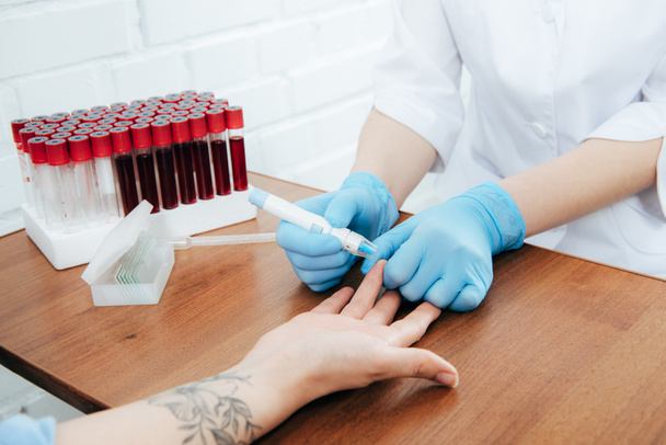
Health care researchers working in life science laboratory. Male research scientist and supervisor preparing and analyzing microscope slides in research lab. Invention of the coronavirus vaccine
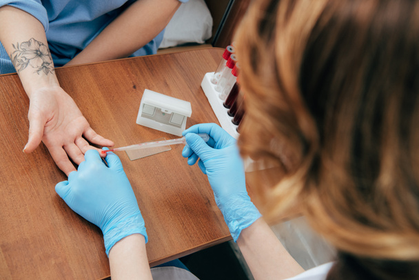
Health care researchers working in life science laboratory. Young researchs preparing and analyzing microscope slides in research lab.

Focused senior life science professional routine screening the microscope slides in the cell laboratory. Lens focus on the researchers face.


Health care researchers working in life science laboratory. Male research scientist and supervisor preparing and analyzing microscope slides in research lab. Invention of the coronavirus vaccine

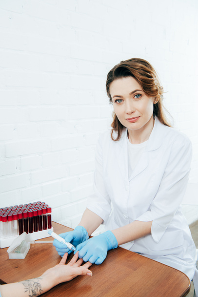
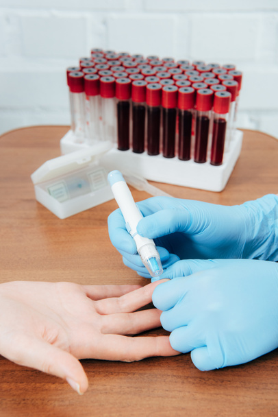
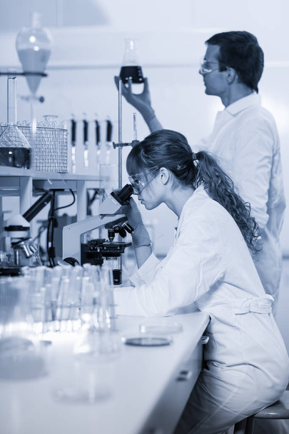
Health care researchers working in life science laboratory. Young female research scientist and senior male supervisor preparing and analyzing microscope slides in research lab.
Health care researchers working in life science laboratory. Young female research scientist and senior male supervisor preparing and analyzing microscope slides in research lab.
Health care researchers working in life science laboratory. Young researchs preparing and analyzing microscope slides in research lab.
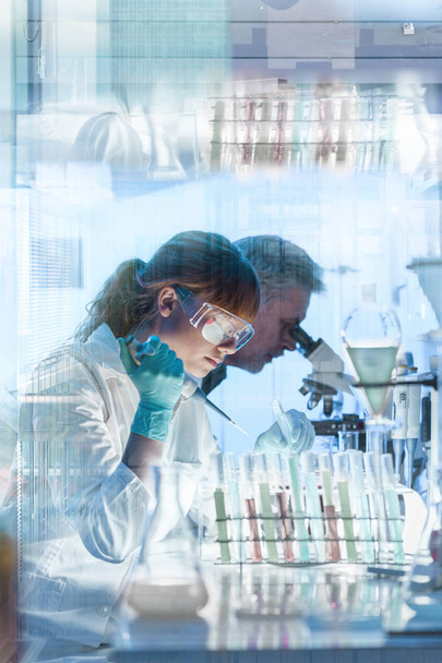
Health care researchers working in life science laboratory. Young female research scientist and senior male supervisor preparing and analyzing microscope slides in research lab.

Health care researchers working in life science laboratory. Male research scientist and supervisor preparing and analyzing microscope slides in research lab. Invention of the coronavirus vaccine

Young female scientist preparing slides with paraffin-embedded sections for pathological analysis .

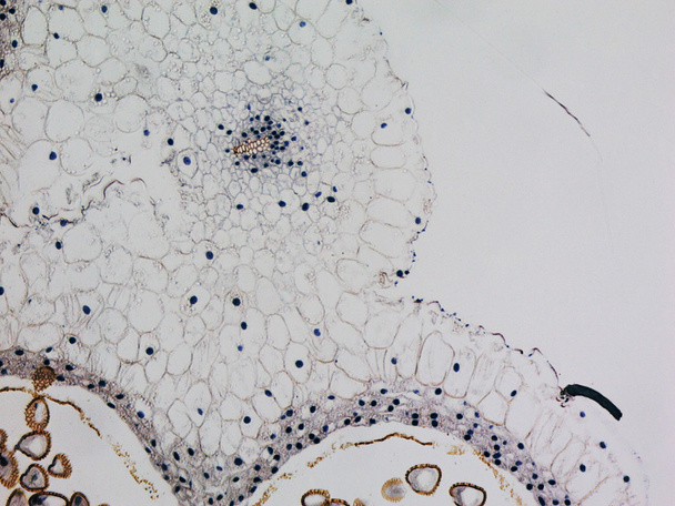
Light micrograph of a cross-section of the bone. Epiphysis of a developing long bone.

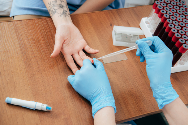
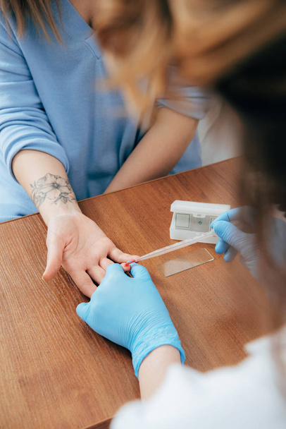
Scientist preparing slides with tissue samples for immunohistochemistry assay in the laboratory. Scientist at the Immunohistochemistry laboratory carry out antigen retrieval on microscope slides with biopsy tissue.

Scientist preparing slides with tissue samples for immunohistochemistry assay in the laboratory. Scientist at the Immunohistochemistry laboratory carry out antigen retrieval on microscope slides with biopsy tissue.

Frozen blocks of paraffin embedded tissue kept cold on ice ready to be cut using a microtome. Paraffin blocks containing biopsy tissue for sectioning. Pathology laboratory. Cancer diagnosis.

Young female scientist preparing slides with paraffin-embedded sections for pathological analysis.
Light photomicrograph of Cotton stem cross section seen through microscope

A young male lab technician looks at slides through a microscope whilst comparing the results to earlier tests.


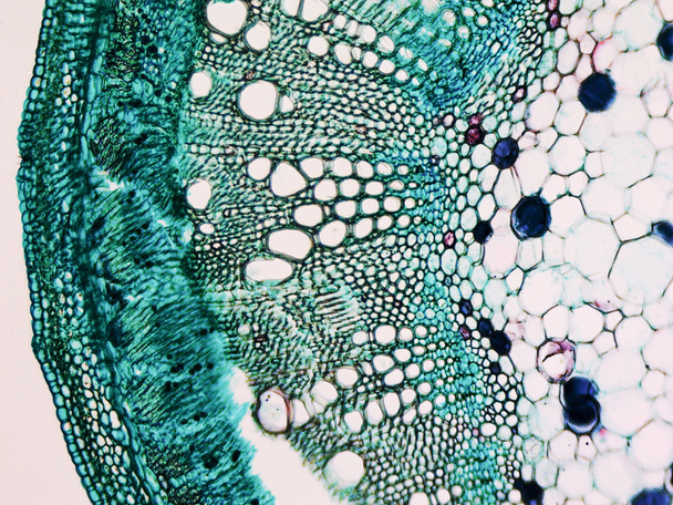
Light photomicrograph of Cucurbita stem cross section seen through microscope

Light photomicrograph of Cucurbita stem cross section seen through microscope

Light photomicrograph of Cucurbita stem cross section seen through microscope

Light photomicrograph of Mulberry seen through microscope

Light photomicrograph of Cucurbita stem cross section seen through microscope

Light photomicrograph of Helianthus stem cross section seen through microscope

Light photomicrograph of Bamboo Stem cross section seen through microscope

Light photomicrograph of tilia stem cross section seen through a microscope

Light photomicrograph of Cucurbita stem cross section seen through microscope

Light photomicrograph of pine tree wood seen through a microscope

Light photomicrograph of Cucurbita stem cross section seen through microscope

Light photomicrograph of Bamboo Stem cross section seen through microscope
Light photomicrograph of Lily anther cross section seen through microscope
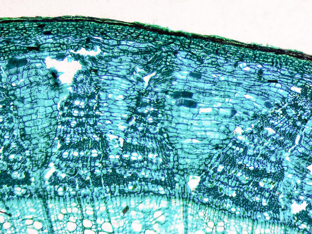
Light photomicrograph of tilia stem cross section seen through a microscope

Light photomicrograph of pine tree wood seen through a microscope

Light photomicrograph of tilia stem cross section seen through a microscope

Light photomicrograph of Cucurbita stem cross section seen through microscope

Light photomicrograph of tilia stem cross section seen through a microscope

Light photomicrograph of tilia stem cross section seen through a microscope

Light photomicrograph of Bamboo Stem cross section seen through microscope

Light photomicrograph of pine tree wood seen through a microscope

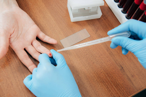
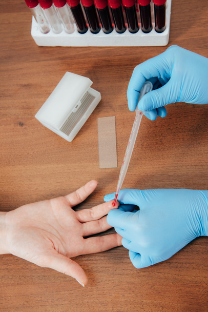
Microscope slide staining tissue biopsy for diagnosis in pathology laboratory. Staining is an auxiliary technique used in microscopy to enhance contrast in the microscopic image.

Light photomicrograph of pine tree wood seen through a microscope

Giant platlet on red blood cells background.

Light photomicrograph of Vicia Faba young root cross section seen through microscope

Light photomicrograph of colour halftone print dots seen through a microscope

Light photomicrograph of tilia stem cross section seen through a microscope

Light photomicrograph of pine tree wood cross section seen through a microscope

Light photomicrograph of pine tree wood cross section seen through a microscope

Light photomicrograph of tilia stem cross section seen through a microscope

Light photomicrograph of Cucurbita stem cross section seen through microscope

Light photomicrograph of tilia stem cross section seen through a microscope

Light photomicrograph of tilia stem cross section seen through a microscope
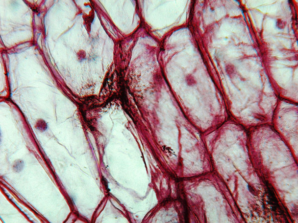
Light photomicrograph of an Onion epidermus cells seen through a microscope

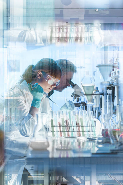
Researchers Health care working in life science laboratory. Young female research scientist preparing and analyzing microscope slides in research lab

Researchers Health care working in life science laboratory. Young female research scientist preparing and analyzing microscope slides in research lab

Light photomicrograph of Mulberry seen through microscope

Light photomicrograph of Vicia Faba young root cross section seen through microscope

Light photomicrograph of tilia stem cross section seen through a microscope

Light photomicrograph of an Onion epidermus cells seen through a microscope

Light photomicrograph of pine tree wood cross section seen through a microscope

Light photomicrograph of an Onion epidermus cells seen through a microscope

Light photomicrograph of Cucurbita stem cross section seen through microscope

Light photomicrograph of Cucurbita stem cross section seen through microscope

Light photomicrograph of pine tree wood cross section seen through a microscope

Light photomicrograph of courgette aka zucchini cells seen through a microscope

Light photomicrograph of pine tree wood cross section seen through a microscope

Light photomicrograph of pine tree wood cross section seen through a microscope

Giant platlet on red blood cells background.

Light photomicrograph of Mitosis of onion root tip cells seen through microscope in black and white

Giant platlet on red blood cells background.

Light photomicrograph of an Onion epidermus cells seen through a microscope

Light photomicrograph of pine tree wood cross section seen through a microscope

Light photomicrograph of Corn root tip cross section seen through microscope
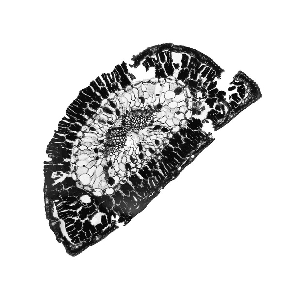
Light photomicrograph of Pine leaf cross section seen through microscope in black and white

Researchers Health care working in life science laboratory. Young female research scientist preparing and analyzing microscope slides in research lab

Light photomicrograph of colour halftone print dots seen through a microscope

Light photomicrograph of Lily ovary cross section seen through microscope

Light photomicrograph of pine tree wood seen through a microscope

Light photomicrograph of pine tree wood seen through a microscope

Light photomicrograph of an Onion epidermus cells seen through a microscope

Light photomicrograph of pine tree wood cross section seen through a microscope

Light photomicrograph of tilia stem cross section seen through a microscope

Light photomicrograph of courgette aka zucchini cells seen through a microscope

Light photomicrograph of Lily ovary cross section seen through microscope
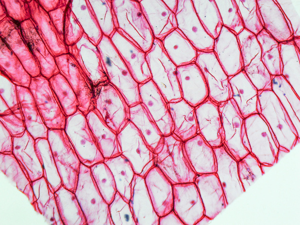
Light photomicrograph of Onion epidermus cells seen through a microscope

Light photomicrograph of pine tree wood cross section seen through a microscope

Light photomicrograph of Bamboo Stem cross section seen through microscope

Microscope slide staining tissue biopsy for diagnosis in pathology laboratory. Staining is an auxiliary technique used in microscopy to enhance contrast in the microscopic image.

Light photomicrograph of Lily anther cross section seen through microscope

Light photomicrograph of Corn root tip cross section seen through microscope

Light photomicrograph of Pine leaf cross section seen through microscope