Social Media


Popular features
Photo

Filters

Stock vector images of Retikulum
Discover royalty-free, professionally-designed vector art of Retikulum for personal and commercial use.


Anatomy of plant cell (Biology Diagram) illustration


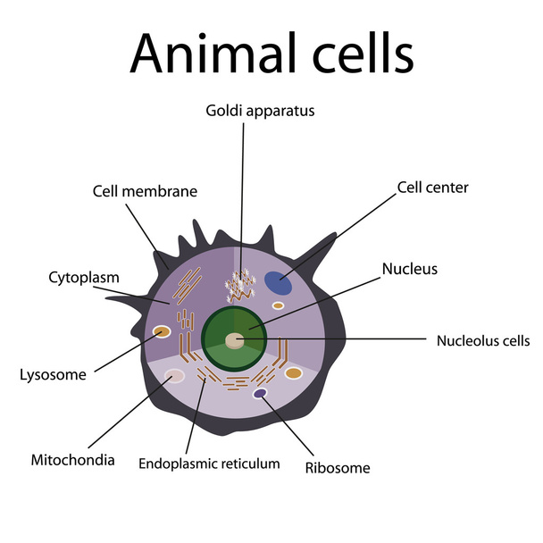
Anatomy of an animal cell. Educational poster


Structure of human cells. Organelles. The core nucleus, endoplasmic reticulum, Golgi apparatus, lysosomes, ribosomes, mitochondria centriole Vector illustration


Endoplasmic reticulum structure. Infographics. Vector illustration on isolated background.


Structure Goblet cells of the intestine. Infographics. Vector illustration on isolated background.


Fibroblast is a dermis cell (vital to the skin's strength and elasticity). Structure of Fibroblast cell.


Animal cell anatomy diagram illustration


Structure of human cells. Organelles. The core nucleus, endoplasmic reticulum, Golgi apparatus, lysosomes, ribosomes, mitochondria, centriole Vector illustration on a black background


Lysosomes of the cell.


Illustration of Endoplasmic reticulum - rough and smooth endoplasmic reticulum.

Illustration showing the natural environment

Enterocyte. Intestinal absorptive cells, are simple columnar epithelial cells which line the inner surface of the small and large intestines.

Organelles of animal cells. Vector illustration for biological, science and educational use

Anatomy of animal cell. Human cell structure. All organelles: Nucleus, Ribosome, Rough endoplasmic reticulum, Golgi apparatus, mitochondrion, cytoplasm, lysosome, Centrosome. Isometric flat vector illustration

Illustration showing the DNA chromosome

Illustration of a plant cell

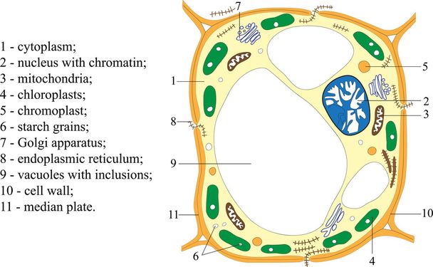
Labelled diagram of the structure of a plant cell

Human or animal cell. cross section. structure of a Eukaryotic cell. Vector diagram for your design, educational, medical, biological and science use

Cell organelles. Structure and anatomy of a animal cell. realistic cell on a white background. Vector illustration. Poster for education

Plant cell anatomy in nature illustration

Reticulum is a caul or coif of network for covering the hair, vintage line drawing or engraving illustration.

Mucous stomach with erosive gastritis. Infographics. Vector illustration on isolated background.
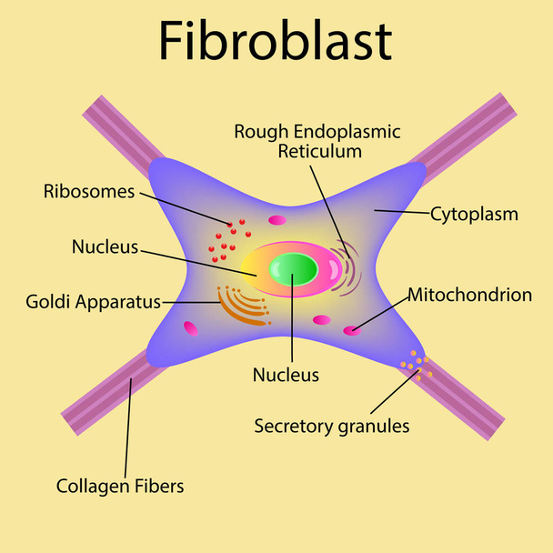
Vector illustration. Fibroblast is a dermis cell. Structure of Fibroblast cell.

Yeast cell scheme for biological lessons art design stock vector illustration for web, for print

Anatomy of nucleus cells.

Anatomy of nucleus cells.

Diagrammatic illustration of stage III ovarian cancer, anatomy of the uterus and ovaries, anatomy of the uterus and ovaries - Translation: Cancer has spread to the lymph nodes or across the pelvic cavity to the peritoneum, large mesentery, or small i

Grate grid pattern. Fiber, wicker interlock mesh design background. Abstract lattice, grill, trellis element. Intersect, cross matrix, array of lines. Abstract geometric texture

Anatomy of plant cell (Biology Diagram) illustration

Plant Cell Anatomy diagram illustration

Mucous stomach with atrophic gastritis. Infographics. Vector illustration on isolated background.

Gastric mucosa in polypous gastritis. Infographics. Vector illustration on isolated background.
The scheme of structure of the plant cell (poster)
The internal structure of an animal cell. Illustration. Isolated on a white background.

Illustration of the daisyheads

Detailed scheme of yeast cell with bud and scar. Yeast cell as basic representation of fungus cell.

Structure of a Golgi complex. Close-up of Golgi apparatus anatomy. Cross section of cell organelle. isometric flat vector illustration

Microscopic structure of cell. Cytoplasm with elements of golgi apparatus and ribosomes accumulation of mitochondria and cytoplasm in vector endoplasmic reticulum.

Cross section of a eukaryotic cell

Fibroblast and Human skin structure (Muscles, Fat cell, Hyaluronic acid, Elastin, Collagen, Fibroblast).

Plant cell isolated on white photo-realistic vector illustration

Anatomy of the Lysosome: Hydrolytic enzymes, Membrane and transport proteins. This organelle use the enzymes to break down and digest food particles, engulfed viruses or bacteria in the cell. Vector diagram for medical use

The structure of the Golgi apparatus. Infographics. Vector illustration on isolated background.

Simplified minimalistic organic cell. Healthy pink microscopic formation of organism that creates organism and participates in biological vector processes.

Cell anatomy. Cell structure and organelles Nucleus, Ribosomes, Endoplasmic reticulum, Golgi apparatus, mitochondrion, cytoplasm, lysosome. Close-up of lipid bilayer cell membrane. Vector poster. Isometric Flat illustration.
Fence Vector Illustration
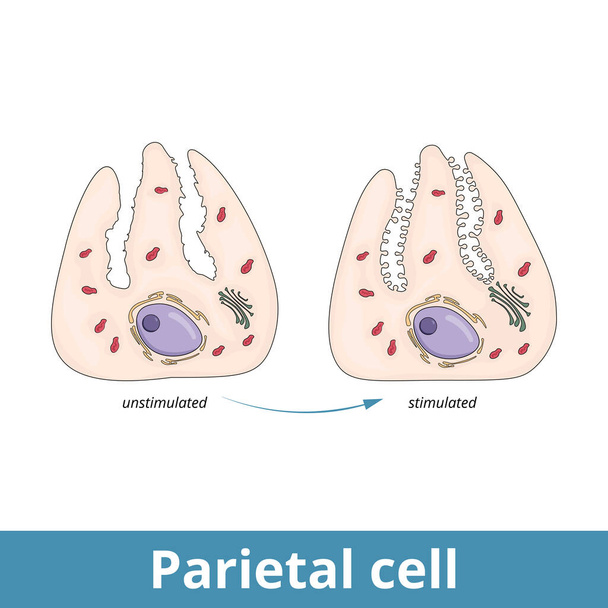
Parietal cell. Epithelial cells in the stomach that secrete hydrochloric acid and intrinsic factor, located in the gastric glands, found in the lining of the fundus and body regions of the stomach.

A caulor coif of network for covering the hair vintage illustration

Anatomy of animal cell (Biology Diagram) illustration
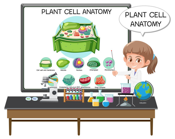
Young scientist explaining the anatomy of the plant cell (Biology Diagram) illustration

Ribosomes are macro-molecular production units.