


Filters

Stock royalty-free photos and images of Optický nerv
Discover unlimited high resolution images of Optický nerv and stock visuals for commercial use.


Human eye anatomy. Inner structure. 3d illustration

Picture of a human eye close up

Superior view of the path and transmission of visual information from the retina.
Anatomy of an eye in section showing lens, retina. .

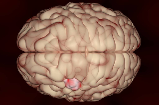
Left and right hemisphere brain areas and midline structures.

Left and right hemisphere brain areas and midline structures.

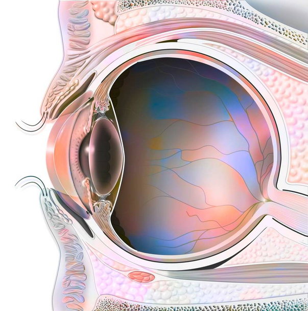
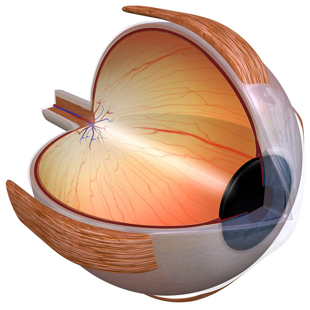
Sagittal view of the eye anatomy showing lens, retina, cornea, iris, choroid. .

Glioblastoma, Brain metastasis,MRI Brain The doctor pointed out the location of the brain tumor on the computer screen

Second step in the functioning of the brain when we fall in love. The signal is sent to the amygdala, as for the perception of danger.

The optic tract: transmission of visual information from the retina to the visual cortex.

Eyeballs with optic nerve and iris

The eye and retina with the vitreous, the internal limiting membrane. .

Text sign showing Glaucoma. Business photo showcasing Eye diseases which result in damage to the optic nerve Vision loss Asymmetrical uneven shaped format pattern object outline multicolour design

Eyeballs with optic nerve and iris

Word writing text Glaucoma. Business photo showcasing Eye diseases which result in damage to the optic nerve Vision loss

Conceptual hand writing showing Glaucoma. Concept meaning Eye diseases which result in damage to the optic nerve Vision loss

Closeup of Human artificial eyes used in hospitals

Text sign showing Glaucoma. Conceptual photo Eye diseases which result in damage to the optic nerve Vision loss.

Handwriting text Glaucoma. Concept meaning Eye diseases which result in damage to the optic nerve Vision loss.

Handwriting text Glaucoma. Conceptual photo Eye diseases which result in damage to the optic nerve Vision loss

Writing note showing Glaucoma. Business photo showcasing Eye diseases which result in damage to the optic nerve Vision loss.

Large moles near the eye close-up. Red large round moles on the woman's face. Inflammation of the tear duct. A woman's reddened eye. Spots on the face and under the eyes in liver diseases

Text caption presenting Glaucoma, Business showcase Eye diseases which result in damage to the optic nerve Vision loss Downloading And Writing Online Content, Abstract Replying To Emails

Woman using eye drop, woman dropping eye lubricant to treat dry eye or allergy sick girl treating eyeball irritation or inflammation sick woman suffering from irritated eye, optical symptoms

World Glaucoma Week is observed every year in March, it is a group of eye conditions that damage the optic nerve, the health of which is vital for good vision. 3D Rendering

Conceptual hand writing showing Glaucoma. Business photo showcasing Eye diseases which result in damage to the optic nerve Vision loss.

Writing displaying text Glaucoma, Business concept Eye diseases which result in damage to the optic nerve Vision loss Abstract Typing A Good Restaurant Review, Ordering Food Online Concept

Conceptual hand writing showing Glaucoma. Business photo text Eye diseases which result in damage to the optic nerve Vision loss.

Text sign showing Glaucoma. Business photo text Eye diseases which result in damage to the optic nerve Vision loss Trendy metallic laptop blank sticky pad coffee cup pen lying vintage table

Glaucoma, general term used to describe a group of eye disorders that damage your optic nerve. Creative text carved and colored on a white stone witn black volcanic sand background

Writing note showing Glaucoma. Business concept for Eye diseases which result in damage to the optic nerve Vision loss

Glaucoma word cloud collage, medical concept backgroun

Conceptual hand writing showing Glaucoma. Business photo showcasing Eye diseases which result in damage to the optic nerve Vision loss.

Hand writing sign Glaucoma, Business showcase Eye diseases which result in damage to the optic nerve Vision loss Building An Unfinished White Jigsaw Pattern Puzzle With Missing Last Piece

Text sign showing Glaucoma, Concept meaning Eye diseases which result in damage to the optic nerve Vision loss Thinking New Bright Ideas Renewing Creativity And Inspiration

Sign displaying Glaucoma, Business showcase Eye diseases which result in damage to the optic nerve Vision loss New Ideas Brainstoming For Maintenance Planning Creative Thinking
Meningioma (brain cancer) tumor in the brain tissue - 3d illustration top view

World Glaucoma day is observed every year on March 12, it is a group of eye conditions that damage the optic nerve, the health of which is vital for good vision. 3D Rendering
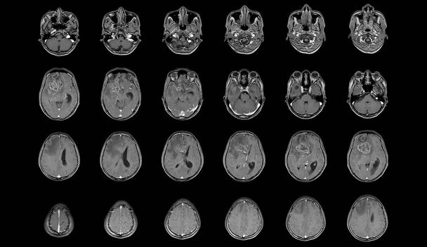
MRI Brain Axial views .to evaluate brain tumor. Glioblastoma, brain metastasis isodensity mass with an ill-defined margin and surrounding edema at the right frontal lobe.
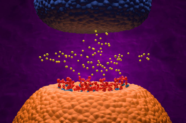
Autoantibodies bond to receptor (achr) blocking the acetylcholine transmitters in Myasthenia gravis (MG) - 3d illustration isometric view

Eye of a cow being dissected is presented - close-up

Eye anatomy and sectional eyelids with lens, retina. .

Ependymoma cancer cells (brain tumor) - isometric view 3d illustration

Glioblastoma multiforme (GBM) brain cancer - closeup view 3d illustration

Blood-brain barrier (BBB) in the human brain - front view 3d illustration

Human Brain Anatomy For Medical Concept 3D Illustration

Human Brain Anatomy For Medical Concept 3D Illustration
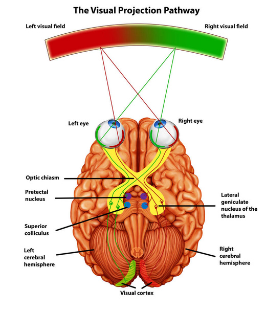
Visual projection pathway illustration
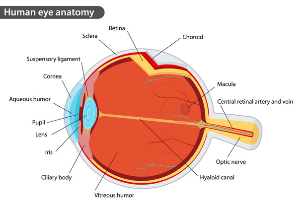
Human eye anatomy, with names

The structure of the eye. Blurred vision in glaucoma. As the eye can see the affected with glaucoma

Abstract optical fiber and high speed internet background. Futuristic computer, internet and technology concept, moving colorful digital glowing lines.

Microscopic view of the neurons. Brain region, optic lobe, drosophila melanogaster neuron. Connections and communication and brain stimulation, 3d rendering

Anatomy of brain system, brainstem, 3d illustration

Anatomy of the eye

Human Brain Anatomy For Medical Concept 3D Illustration

The optic nerve connects each eye to the brain.3D rendering

They may not be painful and you may not see changes in your vision until the disease has become very advanced
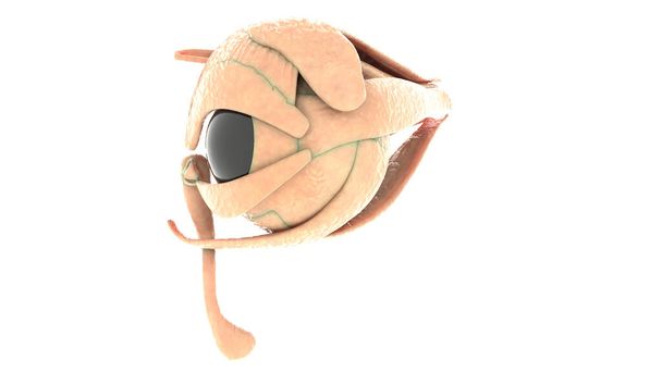
3D rendering of the human eye, quarter view

Human Brain Anatomy For Medical Concept 3D Illustration

Small boy with slit lamp microscope for eye examination.
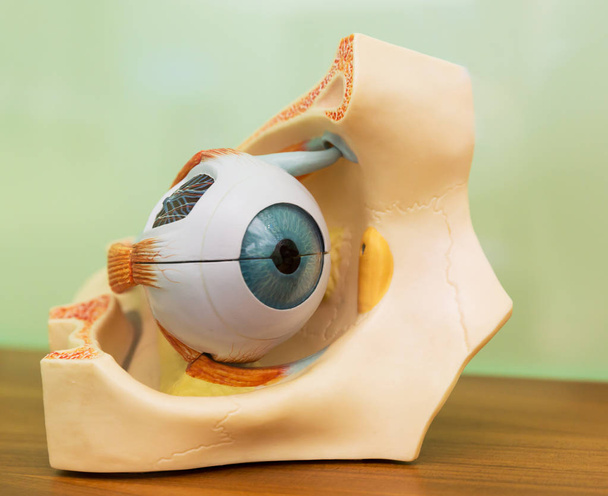
Anatomical plastic model of human eye. Medical stand, eyeball education concept

View inside human eye disorders showing retina, optic nerve and macula Severe age-related macular degeneration.Medical image Retina Abnormal isolated on black background
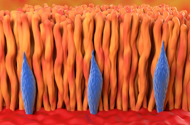
Retina cone and rod in the human eye isometric view 3d illustration
Human Organs Anatomy (Eye). 3D

Ophthalmology diagnosis Glaucoma. Snellen (eye) chart, two bottles of eye drops ( medications) lying on notebook with inscription Glaucoma diagnosis on the desk in ophthalmologist office

Close-up image of an eye looking on the magnifying glass

MRI OF BRAIN AND ORBIT HISTORY: A 50-year-old woman, presented with acute headache and exophthalmos of right eye FINDINGS :The study reveal a well-circumscribed, expansile growth lesion, occupying at right anterior ethmoid and right frontal sinuses.

Human Eye Dissection Anatomy

3d Astigmatism Concept word cloud

World Glaucoma Day written on blackboard by color chalks and stethoscope. Medical concept.
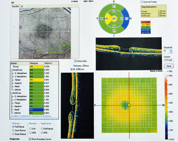
OCT of the eye reveals faint epimacular membrane and full thickness macular hole involving the fovea, surrounding diffuse macular oedema showing few cystoid changes for follow up, selective focus

Anatomical plastic model of human eye close up

Human Organs Anatomy (Eye). 3D

Conceptual hand writing showing Glaucoma. Concept meaning Eye diseases which result in damage to the optic nerve Vision loss

Close shot of the human eye. The eye of a man.

Word writing text Glaucoma. Business photo showcasing Eye diseases which result in damage to the optic nerve Vision loss

Long eyelashes. Mascara. macro

Out of focus patients are examining their eyesight with a Optical Coherence Tomography in a screening room for patients at risk of diabetes.Medical healthcare concept

Normal eye retina, ophthalmoscope view, scientific 3D illustration showing optic disk, blood vessels, macula and fovea
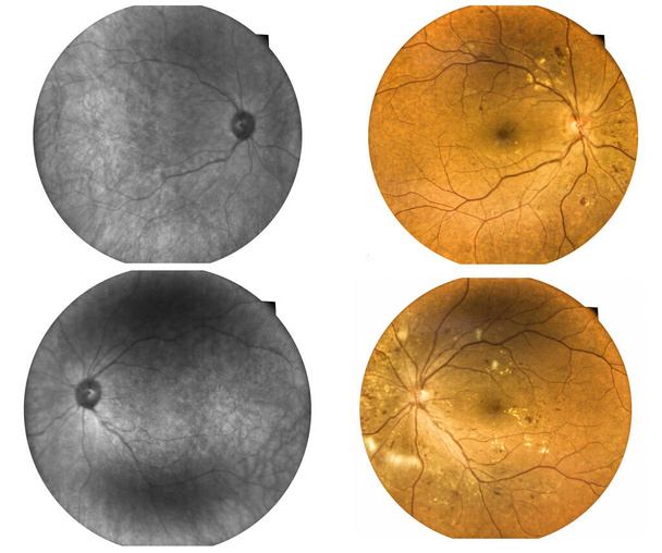
Fundus photography Madical Retina IR and color Abnormal isolated on white background.Retina of diabetes check up medical healthcare concept.

The Eyeball, vintage engraved illustration

Close-up image of a man's left eye looking at the camera

Close-up of a man's eye looking at the camera

The close up shot of human eye. The human eye is a paired sense organ that reacts to light and allows vision.

Human eye nerve under the microscope view. Histological for human physiology.

Close up image of the left eye of a man looking at the side

Close shot of the human eye. The eye of a man.
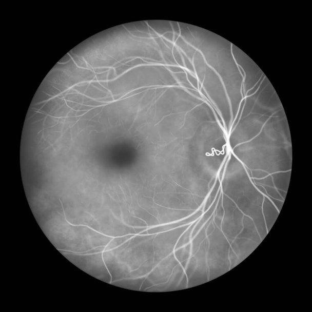
A prepapillary vascular loop on the retina, as observed during ophthalmoscopy in fluorescein angiogram, an illustration showcasing the looping blood vessels around the optic disc.

Cataract word written under brown torn paper. Eyes healthcare concept.

Left eye of a white male close-up, soft focus.

Eye retina, scientific illustration showing optic disk, blood vessels, macula and fovea, ophthalmoscope view, fluorescein angiography
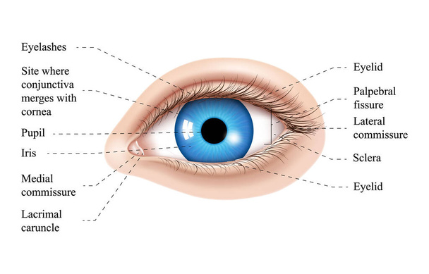
Human eye anatomy illustration. Parts of the eye, labeled vector illustration diagram. Eyelid, eyelashes, pupil, lacrimal gland and other anatomical parts. Realistic 3d vector isolated

Retina in blastomycosis (infection caused by fungi Blastomyces dermatitidis) as seen during ophthalmoscopy. 3D illustration showing scattered yellow choroidal infiltrates and a choroidal mass lesion.

A prepapillary vascular loop on the retina, as observed during ophthalmoscopy, 3D illustration showcasing the looping blood vessels around the optic disc.

Retina in Presumed Ocular Histoplasmosis Syndrome as seen in fluorescein angiography, illustration shows punched-out atrophic and pigmented chorioretinal scars (histo spots) and peripapillary scarring

Retina surface (cones and rods) in the human eye closeup view 3d illustration

OCT of the eye reveals faint epimacular membrane and full thickness macular hole involving the fovea, surrounding diffuse macular oedema showing few cystoid changes for follow up, selective focus

OCT of the eye reveals faint epimacular membrane and full thickness macular hole involving the fovea, surrounding diffuse macular oedema showing few cystoid changes for follow up, selective focus

Retina human under microscope.