


Filters

Stock royalty-free photos and images of Metastaz
Discover unlimited high resolution images of Metastaz and stock visuals for commercial use.


An illustration depicting Cancer Cells in the crosshairs, related to cancer treatment.


Oncology - Inscription on Red Road Sign on Sky Background.

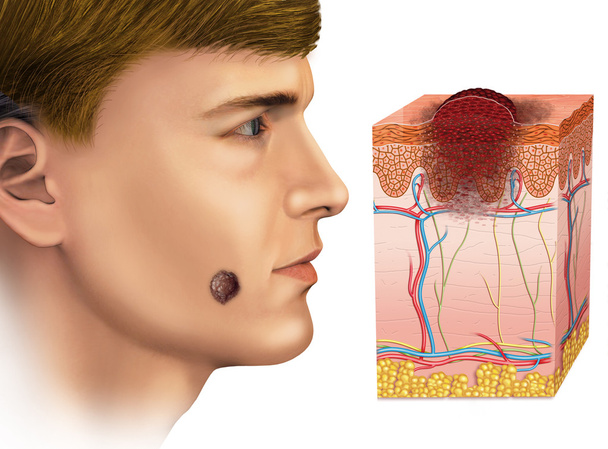
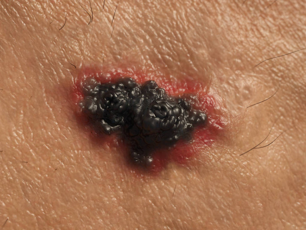
Melanoma in the face


Bone cancer word cloud concept on white background.


Soft tissue sarcoma word cloud and hand with marker concept on white background.
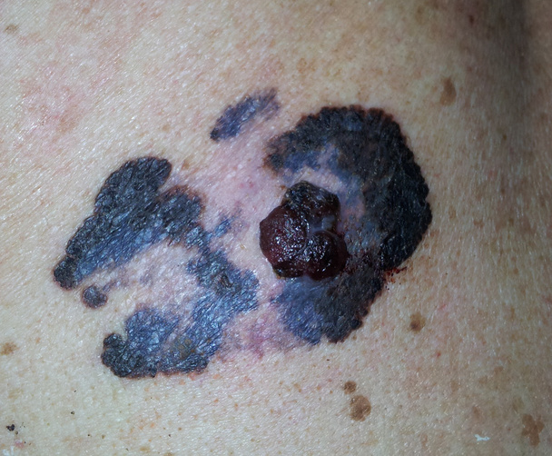
Malignat melanoma, cancer of skin
Melanoma or skin cancer. layers of the human skin.
3d computer illustration of a dendritic cell. They areantigen-presenting cells of the immune system. Their main function is to process antigen material and present it on the cell surface to the T cells of the immune system. They are messengers betwe

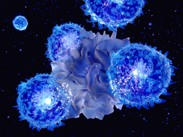
Dendritic cells present antigens (green) to lymphocytes through their membran bound MHC-molecules (violet). CD4 molecules (light blue) bind to other portions of the MHC, strengthening the interaction.

Cancer cells express PD-L1 (orange) proteins on their surface to trick the immune system. The interaction of PD-L1 with PD-1 of T-cells leads to a down-regulation of T-cells. The antibody (yellow) blocks this interaction.
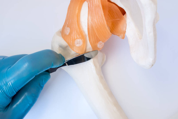
Orthopedic or trauma surgery concept photo. Surgeon holds surgical scalpel in hand, dressed in glove, over femur bone near hip joint, symbolizing operation of orthopedic surgery of bones and joints

Liver cancer concept photo. Anatomical shape of liver lies near letters composing word cancer surrounded by set of tests, analysis, drugs, MRI and stethoscope. Diagnosis treatment of cancerous liver

Lymphoma concept. Purple ribbon in the hand isolated

Skin Cancer - Medical Concept on Blue Background with Blurred Text and Composition of Eyeglasses. 3D Rendering.

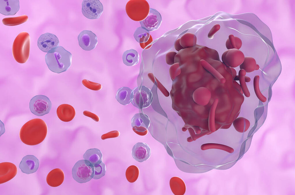
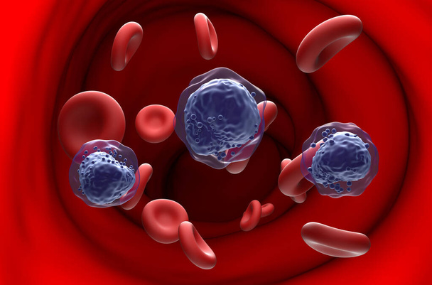
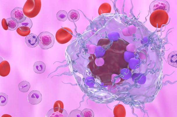
Primary myelofibrosis (PMF) cells in blood flow - closeup view 3d illustration
Meningioma (brain cancer) tumor in the brain tissue - 3d illustration closeup view
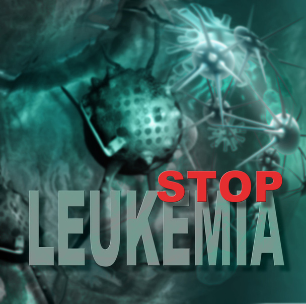
Leukemia cells and words made in 3d software

An illustration depicting cancerous growths on the lungs
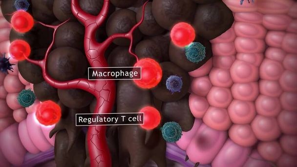
An illustration related to cancer cells and the environments that they spread as depicted in this stylized view from within the body.
Auer rods (or Auer bodies) in acute Hypergranular Promyelocytic Leukemia (APL) - 3d illustration closeup view
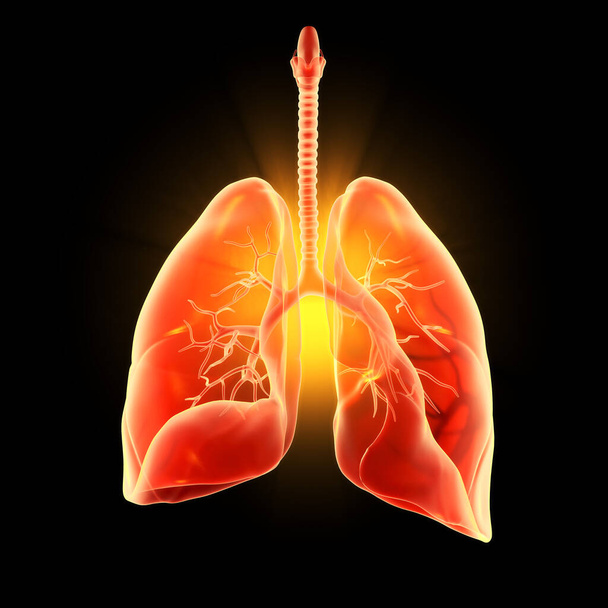
Illustration showing highlighted human lungs, pneumonia, 3D illustration
CAR T cell therapy in Multiple myeloma (MM) - isometric view 3d illustration

Prostate cancer symptoms, a small walnut-shaped gland in men that produces the seminal fluid that nourishes and transports sperm
Primary myelofibrosis (PMF) cells in blood flow - isometric view 3d illustration
Acute myeloid leukemia (AML) cells in blood flow - section view 3d illustration
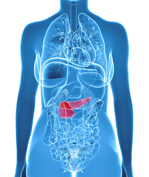
3d illustration with highlighted pancreas
Human Skin cancer. Illustration
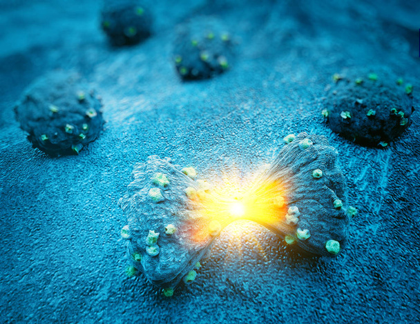
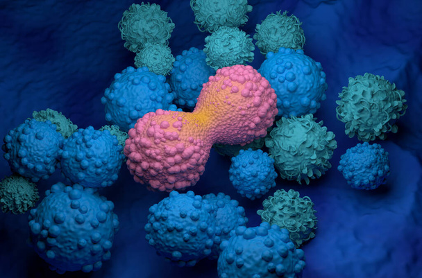
Tumor cell in the moment that divides, 3d illustration

Cancer diagnosis

Metastases. Medical Report with Composition of Medicaments - Red Pills, Injections and Syringe. Selective Focus

Uterus cancer concept. Anatomical shape of uterus with ovaries lies near word cancer surrounded by set of tests, analysis, drugs, MRI and stethoscope. Diagnosis treatment of ovaries or uterus cancer

Close up of leaf change color green yellow red

3D illustration - cancer cell with high details under microscope

Man with blue ribbon, closeup. Colon cancer concept

Man with blue ribbon, closeup. Colon cancer concept
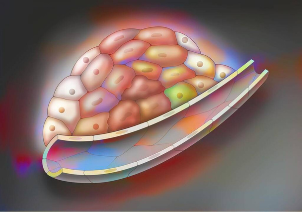
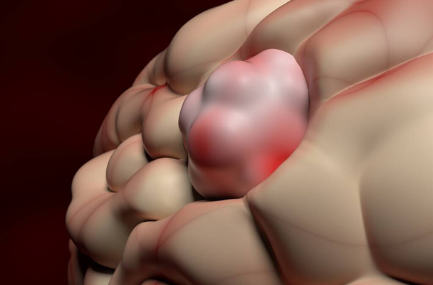
Cancerous tumor, stage 2: Cancer cells have multiplied and transformed.

Liver cancer word cloud and hand with marker concept on white background.

Multiple myeloma word hand sphere cloud concept on white background.

Hodgkin lymphoma word cloud and hand with marker concept on white background.

Skin cancer (non-melanoma) word hand sphere cloud concept on white background.

Oral cancer word cloud and hand with marker concept on white background.

Spinal cancer word cloud and hand with marker concept on white background.

Pancreatic cancer word cloud and hand with marker concept on white background.

Gallbladder cancer word cloud concept on white background.

Spinal cancer word cloud concept on white background.
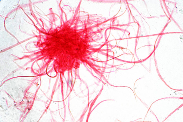
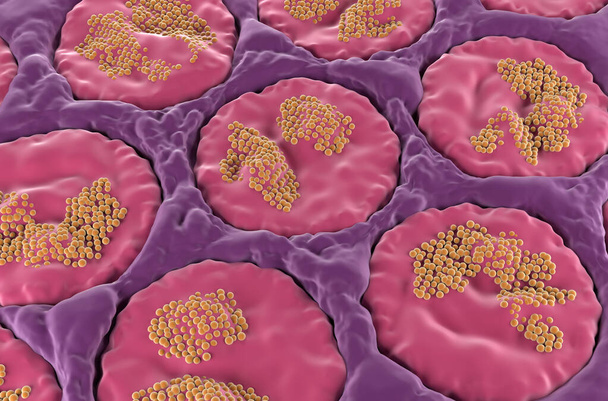
Cancer Cell in human under the microscope view. Medical science background concept.
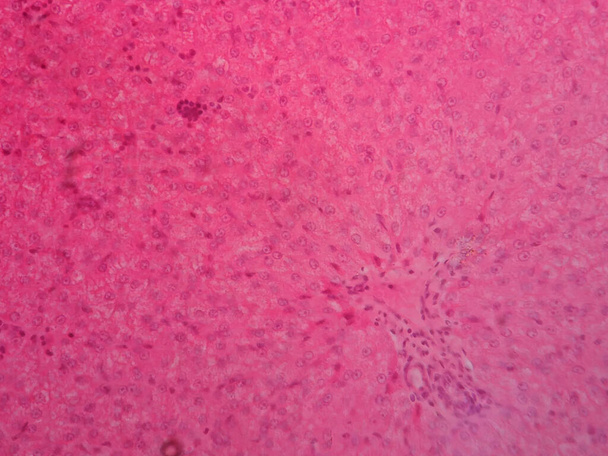
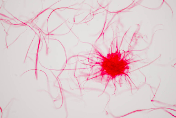
Human liver tissue under microscope view
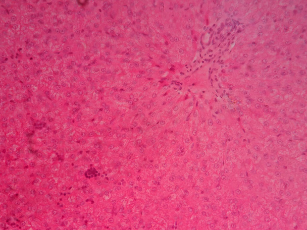
Human liver tissue under microscope view

Human liver tissue under microscope view
Cancer Cell in human under the microscope view. Medical science background concept.

KRAS G12C mutation in non-small cell lung cancer (NSCLC) - isometric view 3d illustration
Salivary gland surface (sublingual) in the human mouth - isometric view 3d illustration
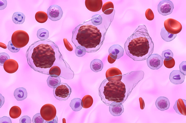
Metastatic neuroendocrine tumor cell in the blood flow - 3d illustration closeup view

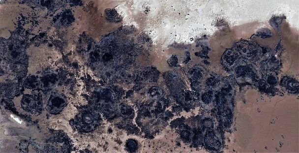
Earth metastasis, black gold, polluted desert sand, tribute to Pollock, vertical abstract photography of the deserts of Africa from the air, aerial view, abstract expressionism, abstract naturalism.
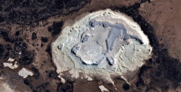
Earth metastasis, black gold, polluted desert sand, tribute to Pollock, abstract photography of the deserts of Africa from the air, aerial view, abstract expressionism, contemporary art,
Earth metastasis, black gold, polluted desert sand, tribute to Pollock, abstract photography of the deserts of Africa from the air, aerial view, abstract expressionism, contemporary art,

Earth metastasis, black gold, polluted desert sand, tribute to Pollock, abstract photography of the deserts of Africa from the air, aerial view, abstract expressionism, contemporary art,

Earth metastasis, black gold, polluted desert sand, tribute to Pollock, abstract photography of the deserts of Africa from the air, aerial view, abstract expressionism, contemporary art,
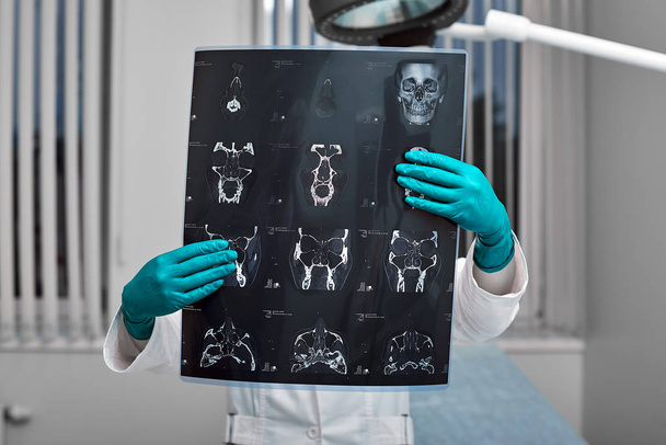
Doctor attentively examines the MRI scan of the patient
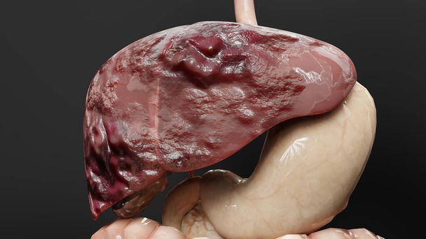
Digestive Organ Liver Cancer, Hepatitis cirrhosis malignant tumor, Hepatic failure, growing cells, duplicating, Fibrosis, Cirrhosis, Hepatocellular carcinoma, Virus infected liver, Human, 3d render

Yellow note paper with word thyroid cancer on cork board background with copy space

Acute myeloid leukemia (AML) cells in blood flow - isometric view 3d illustration

Multiple myeloma cells cluster in the blood flow - isometric view 3d illustration

Wood alphabet letter in word ovarian cancer on wood background

Testicular cancer, testicular seminoma, medical 3D illustration and light micrograph. Malignant tumor of the testis

Bladder cancer is a type of cancer that originates in the urothelial cells of the bladder. 3D rendering

The Bladder Cancer, Stage IV 3D rendering
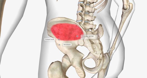
Trabecular or spongy bone is found in the center of the bone. 3D rendering

Digestive Organ Liver Cancer, Hepatitis cirrhosis malignant tumor, Hepatic failure, growing cells, duplicating, Fibrosis, Cirrhosis, Hepatocellular carcinoma, Virus infected liver, Human, 3d render

Cancer cell growth uncontrollably over tissue, Tumor infection cells and spreading, Invasive inflammation metastasis cancerous. reproduce by duplicating, cells expanding, Melanoma Cancer, 3d render

Yellow note paper with word brain cancer on cork board background with copy space

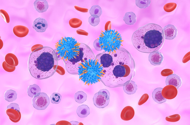
Monoclonal antibody treatment in Diffuse large B-cell lymphoma (DLBCL) - closeup view 3d illustration

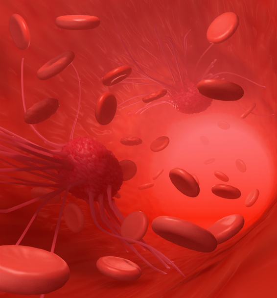
3d rendering of close up view of blood vessel and cancer cell

3d rendering of cancer requires a blood supply to deliver the nutrients and oxygen it needs to grow and survive

Throat cancer word cloud concept on black background.

Vulvar cancer word cloud concept on white background.

Vaginal cancer word cloud concept on white background.

Human prostate medical model with a cross section of the inner organ with red and blue arteries and adrenal gland as a health care and medical of the anatomy system.

KRAS G12C mutation in non-small cell lung cancer (NSCLC) - closeup view 3d illustration

Handwritten Diagnosis Skin Cancer in the Differential Diagnoses. Medicament Composition of Red Pills, Blister of Pills and Bottle of Tablets. 3D.
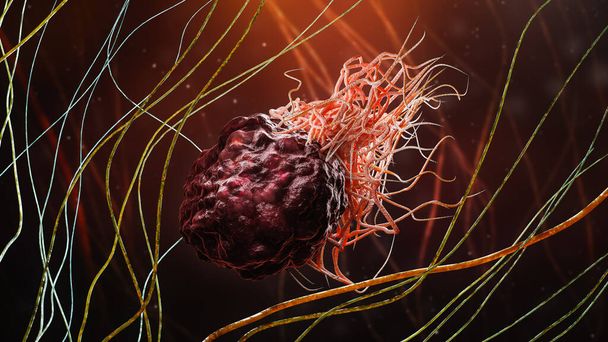
Cancer or tumor cell within fibrous tissue close-up 3D rendering illustration. Carcinoma, lymphoma, oncology, medicine, science, microbiology, cancerous pathology, health concepts.

Immunologically active proteins on a T-cell. TCR (blue), CD-4 (light blue), CD-28 (dark blue), PD-1 (magenta), CTLA-4 (violet), Ca-channel (dark violet). The T-cell receptor, CD-4 and CD-28 activate T-cells, while PD-1 and CTLA-4 inhibit the activat

T-cell receptor in complex with the MHC class II-peptide complex. The antigen (light green) is a peptide from a tumor cell, bacteria or virus. Complex embedded in the membranes. 3D-Rendering. Illustration

Cancer cells, 3d illustration

All blood cells manufactured by stem cells, forming the blood system, medical illustration

Ovarian cancer cell variations - closeup view 3d illustration

Handwritten Diagnosis Cancer in the Extract From the History of Disease. Medicaments Composition of Heap of Pills, Blister of Pills and Bottle of Tablets. 3D Render.
Dividing breast cancer cell isometric view 3d illustration

Stomach Cancer - Gastric Cancer - Cancer that Develops from the Lining of the Stomach - Conceptual Illustration

Acute myeloid leukemia (AML) cells in blood flow - isometric view 3d illustration

3d medical illustration of skin cancer: Squamous cell carcinoma, basal cell cancer

Highlighted carcinoma in right lung, 3D illustration

Lung Cancer - Printed Diagnosis with Blurred Text. On Background of Medicaments Composition - Red Pills, Injections and Syringe
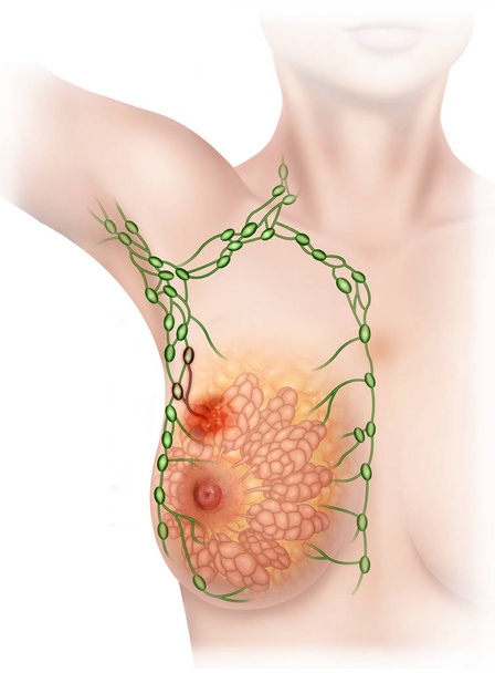
Descriptive illustration of the breasts of a woman in which the lymph and lymphatic system affected by malignant cancer can be seen.

Leukemia cells and words made in 3d software