


Filters

Stock royalty-free photos and images of Microscopy
Discover unlimited high resolution images of Microscopy and stock visuals for commercial use.
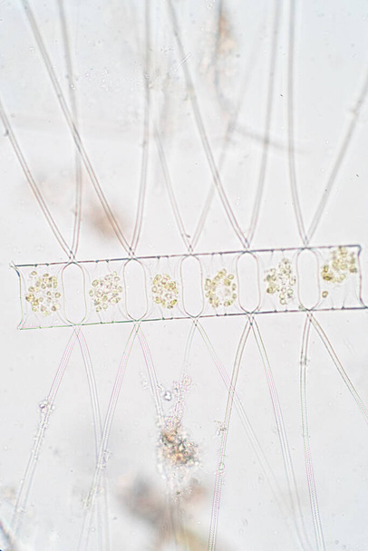
Chaetoceros is the largest genus of marine planktonic diatoms, phytoplankton
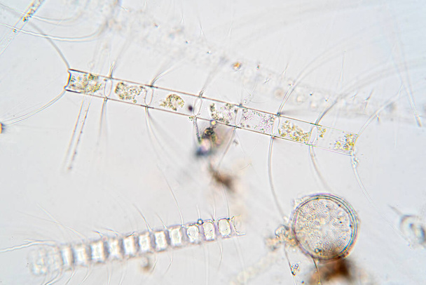
Chaetoceros is marine planktonic diatoms under the microscope view.
Macro shot of microcrystals from Methylparaben in polarized light

Microscopic photo of cannabis. Mautoflowering Lemon Hazre and Purple lemon

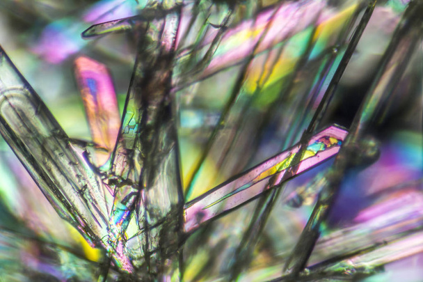
Macro shot of growing microcrystals of soda lye in polarized light

Close up parasite in stool exam.

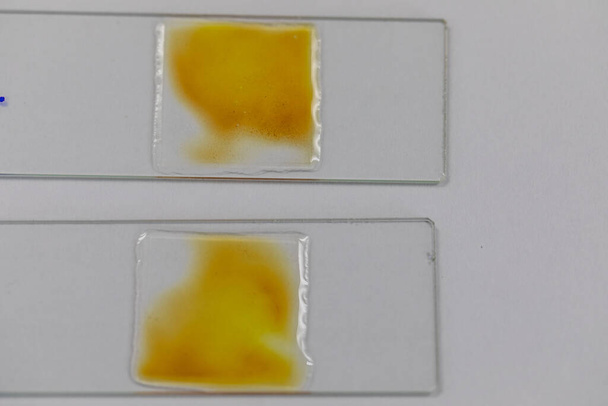
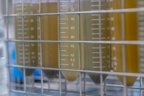
System for detection of inhibitors and antibiotics in animal milk. Milk antibiotic test. The veterinarian is doing a test in the lab.
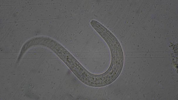
Strongyloides stercoralis larva in stool exam.Parasite in human.
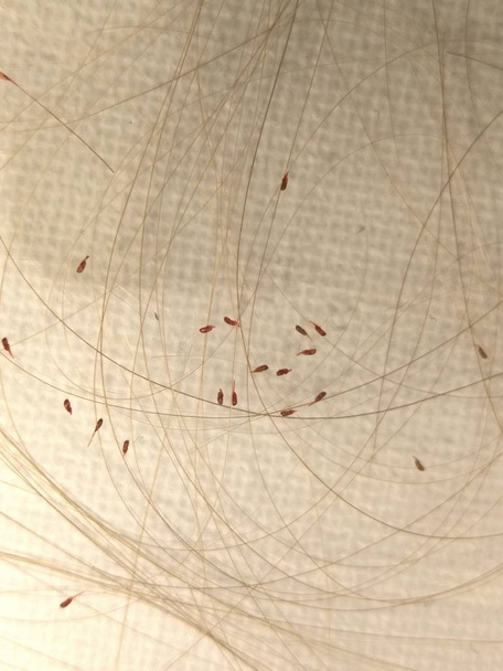
Head lice (louses) isolated

Cell Division and Cell Cycle under the microscope.

Kidney cancer, light micrograph, photo under microscope. High magnification
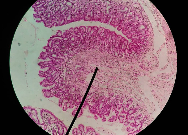
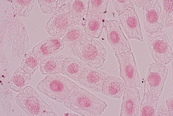
Root tip of Onion and Mitosis cell in the Root tip for education

Mitosis cell in the Root tip of Onion under a microscope.

Microscopic shot showing some microcrystals in warm ambiance

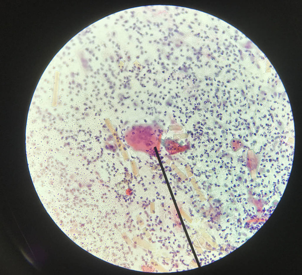
Finding parasites in feces in laboratory.

Macro shot of growing microcrystals of soda lye in polarized light
Cells in reproductive female cytology and histology education concept.

System for detection of inhibitors and antibiotics in animal milk. Milk antibiotic test. The veterinarian is doing a test in the lab.

Root tip of Onion and Mitosis cell in the Root tip of Onion under a microscope.
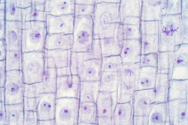
Root tip of Onion and Mitosis cell in the Root tip for education

Cell structure Hydrilla, view of the leaf surface showing plant cells under the microscope for classroom education.

Micro-photo of geological thin section under cross-polarized light. (Oolitic Limestone).

3D illustration of abstract macro render with shallow dept of field. Macro city

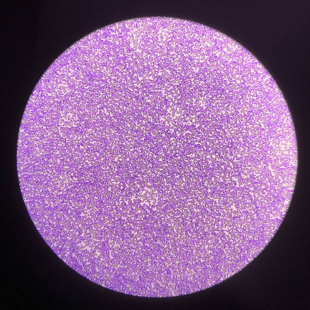
Microscopic shot showing colorful microcrystals in polarised light

Macro shot of growing microcrystals of soda lye in polarized light

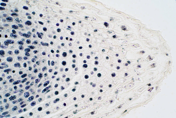
Education anatomy and Histological sample of Human under the microscope.
Finding parasites in feces in laboratory.
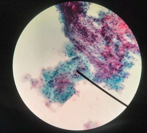
Cells in reproductive female cytology and histology education concept.

Kidney cancer, light micrograph, photo under microscope. High magnification
Cells in reproductive female cytology and histology education concept.

Seeding onion roots to study mitosis cells in Laboratory.

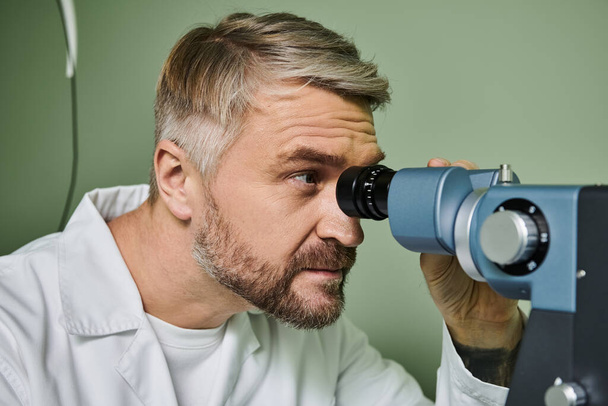
Microscope and Medical laboratory, equipment,Scientific and healthcare research, Scientific microscope lens on blue background, A microscope is an instrument used to see too small objects
Finding parasites in feces in laboratory.

Egg parasite in stool exam laboratory concept.

Root tip of Onion and Mitosis cell in the Root tip of Onion under a microscope.
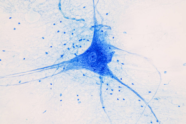
Education Spinal cord and Motor Neuron under the microscope in Lab.
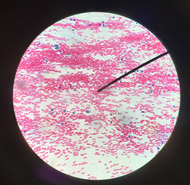
Blood smear for hematology microscopic examination

3D illustration of abstract macro render with shallow dept of field. Macro city
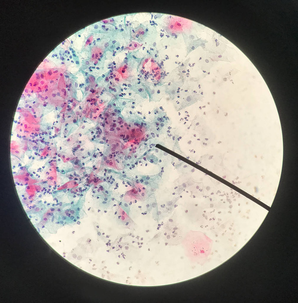
Cells in reproductive female cytology and histology education concept.

Root tip of Onion and Mitosis cell in the Root tip for education

Realistic microscope - 3d Render. Chemistry, pharmaceutical instrument, microbiology magnifying tool. Symbol of science, chemistry and exploration.
Close up human cells with microscope in cytology lab.
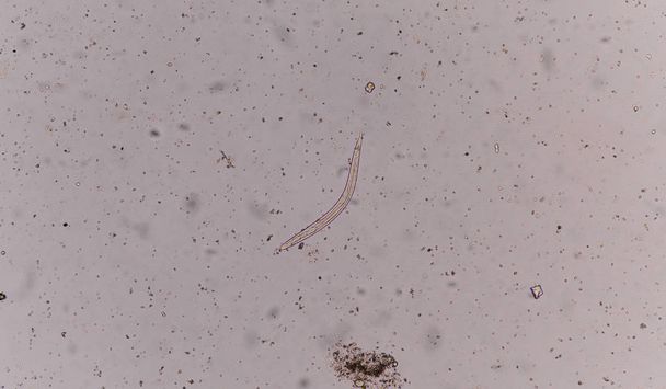
Strongyloides stercoralis (threadworm) in stool, analyzer by microscope

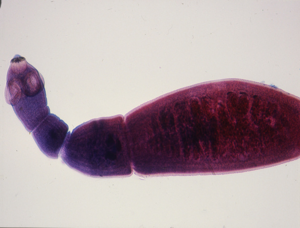
Liver fluke Fasciola hepatica, a parasite of sheep, cattle and humans isolated on white background, light micrograph

Crystallized liquid crystal under polarized light microscope forming a rainbow texture. Abstract squares filled with rainbow colors.

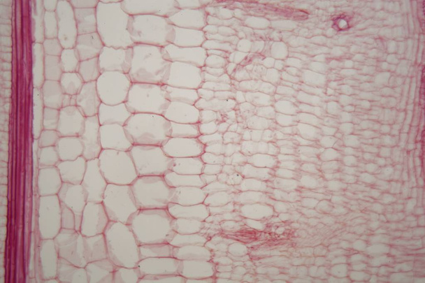
Cross section - Xylem is a type of tissue in vascular plants that transports water and some nutrients. Scientific research. Plant tissue Structure.

Finding parasites in feces in laboratory.

Strongyloides stercoralis (threadworm) in stool, analyzer by microscope

Macro shot of growing microcrystals of soda lye in polarized light

Microscope objective lenses with a prepared slide

Root tip of Onion and Mitosis cell in the Root tip of Onion under a microscope.

Finding parasites in feces in laboratory.

3D illustration of abstract macro render with shallow dept of field. Macro city
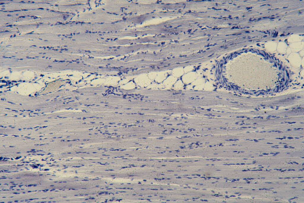
Human skeletal muscle cells under the microscope

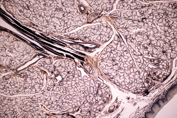
Histology of human compact bone tissue under microscope view for education, muscle bone connection and connective tissue
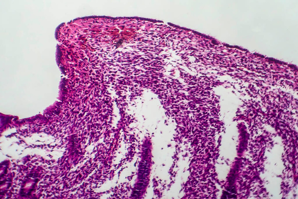
Endometrial hyperplasia, light micrograph, photo under microscope

Struvites at the microscope. Urine sample from a dog who urinate
Finding parasites in feces in laboratory.

Anodonta gills ciliated epithelium under the microscope - Abstract pink and purple color on white background.

Tubular atrophy, light micrograph, photo under microscope. High magnification
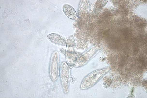
Paramecium is a genus of unicellular ciliated protozoa, Paramecia are widespread in freshwater, brackish, and marine environments and are often very abundant in stagnant basins and ponds.
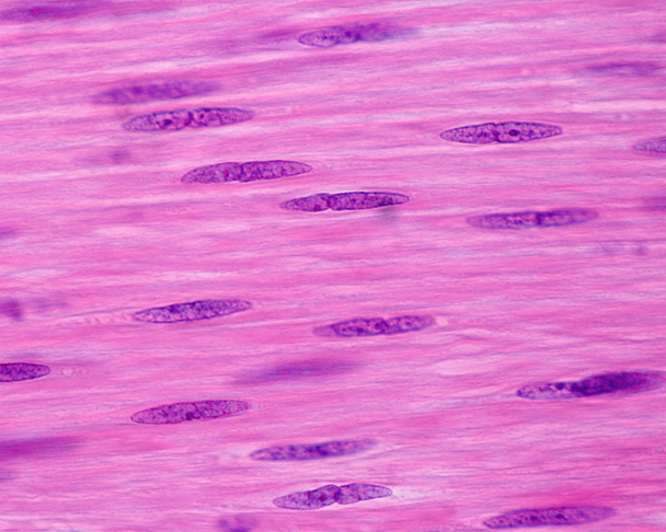
Nuclei of smooth muscle cells. These cells show a very elongated fusiform nucleus which contains small nucleoli. The small incisures in the nucleus surface are due to a contraction of the smooth muscle fiber.

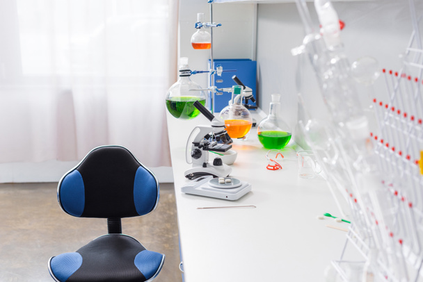
Closeup modern microscope in a lab
Mitosis cell in the Root tip of Onion under a microscope.

Root tip of Onion and Mitosis cell in the Root tip for education

Seeding onion roots to study mitosis cells in Laboratory.
Leukemia blood picture find with microscope 100X.

Microscopic shot showing colorful microcrystals in polarised light

Microscopic detail of some dried egg white in dark back

Background with viruses. Adeno-associated virus serotype 1. Virus is used for gene therapy. 3D illustration
Characteristics of anatomy and Histological sample Striated (Skeletal) muscle of mammal Tissue under the microscope.

Thyroid cancer, 3D illustration showing tumor inside thyroid gland and closeup view of cancer cells

Uterine cancer, 3D illustration and light micrograph showing malignant tumor in the female uterus
Blood smear for hematology microscopic examination

Close up stoma plants cells find with microscope.

Elastic cartilage of human outer ear, light micrograph
Spinal cord, cross-section. 3D illustration which shows the white and the grey matter with dorsal and ventral horns
Tapeworm with segments and suction cups in high magnification
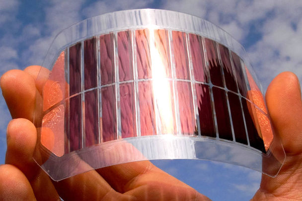
Flexible solar cells from ruthenium. Energy efficiency products

Cross section - Xylem is a type of tissue in vascular plants that transports water and some nutrients. Scientific research. Plant tissue Structure.

Macro shot of growing microcrystals of soda lye in polarized light

Old Lens scope on wood table background

Plant cell under the microscope view for education.

Cell structure Hydrilla, view of the leaf surface showing plant cells under the microscope for classroom education.

Root tip of Onion and Mitosis cell in the Root tip of Onion under a microscope.

Root tip of Onion and Mitosis cell in the Root tip of Onion under a microscope.
Plant stem with sieve cells under the microscope.

Endometrial adenocarcinoma, light micrograph, photo under microscope

Root tip of Onion and Mitosis cell in the Root tip of Onion under a microscope.

Root tip of Onion and Mitosis cell in the Root tip of Onion under a microscope.

Education Spinal cord and Motor Neuron under the microscope in Lab.

Moderate Dysmorphic red blood cells in urine sediment.

Tuber with starch grains across 100x

Researcher or scientist or PHD student working in a biotechnology laboratory sampling DNA in a test tube with centrifuge

Microscopic detail of some dried egg white in dark back

Macro shot of growing microcrystals of soda lye in polarized light

Microscopic shot showing some microcrystals in dried dyestuff

Strongyloides stercoralis larva in stool exam.Parasite in human.