


unnaugan

Chemical formula, structural formula and 3D ball-and-stick model of synthetic antibiotic linezolid, white background

Chemical formula, structural formula and 3D ball-and-stick model of the anticancer drug pemetrexed, white background

Chemical formula, structural formula and 3D ball-and-stick model of etoricoxib, a selective COX-2 inhibitor, white background

Chemical formula, skeletal formula and 3D ball-and-stick model of adenosine triphosphate (ATP), white background

Structure of the brain derived neurotrophic factor (green) - neurotrophin 4 (brown) heterodimer, 3D cartoon and Gaussian surface models, white background

Bovine mitochondrial ATP synthase, 3D Gaussian surface model isolated, chain id color scheme, putative membrane shown, PDB 5ara, white background

Chemical formula, structural formula and 3D ball-and-stick model of firocoxib, a COX-2 inhibitor for veterinary use

Chemical formula, structural formula and 3D ball-and-stick model of celecoxib, a COX-2 inhibitor, white background

Chemical formula, skeletal formula, and 3D ball-and-stick model of nicotinamide adenine dinucleotide, white background

Bovine mitochondrial ATP synthase, 3D ribbon model, black background, isolated

Chemical formula, structural formula and 3D ball-and-stick model of antibiotic cloxacillin, white background

Chemical formula, structural formula and 3D ball-and-stick model of antibiotic methicillin, white background

Chemical formula, skeletal formula and 3D ball-and-stick model of cyclic adenosine monophosphate (cAMP), white background

Chemical formula, skeletal formula, and 3D ball-and-stick model of chemotherapeutic drug clofarabine, white background

Chemical formula, structural formula and 3D ball-and-stick model of the anticancer drug mercaptopurine, white background

Chemical formula, structural formula and 3D ball-and-stick model of pyrazinamide used for treatment of tuberculosis, white background

Chemical formula, structural formula and 3D ball-and-stick model of antibiotic amikacin, white background

Chemical formula, structural formula and 3D ball-and-stick model of isoniazid used for treatment of tuberculosis, white background

Chemical formula, structural formula and 3D ball-and-stick model of antibiotic spectinomycin used for treatment of gonorrhea, white background

Structure of tumor necrosis factor alpha (TNF1) homotrimer. 3D cartoon and Gaussian surface model, white background.

Structure of human interleukin-11, 3D cartoon model isolated, white background

Structure of the extracellular segment of human TRKA in complex with nerve growth factor, 3D cartoon and Gaussian surface models, white background
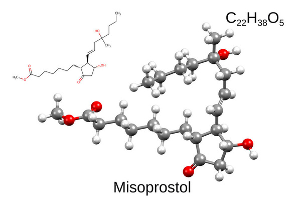
Chemical formula, structural formula and 3D ball-and-stick model of misoprostol, a synthetic prostaglandin medication, white background

A 3D cartoon model of the human CD4 glycoprotein, white background

Chemical formula, skeletal formula and 3D ball-and-stick model of a serotonin A receptor agonist, xaliproden, white background

Chemical formula, skeletal formula and 3D ball-and-stick model of a glutamate antagonist, riluzole, white background

Echinops (Echinops spinosissimus) with fully formed inflorescences before blooming
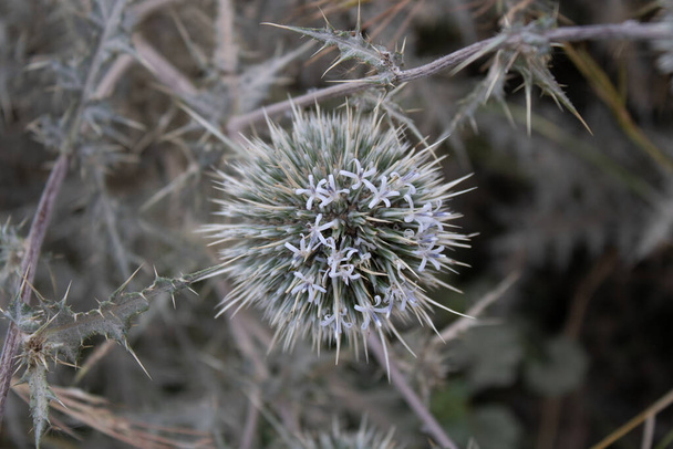
A close-up of a partially blooming echinops (Echinops spinosissimus) inflorescence

Chemical formula, structural formula and 3D ball-and-stick model of mifepristone, also known as RU-486, a medication used to bring about a medical abortion during pregnancy, white background

Structure of human interleukin-21, 3D cartoon model isolated with the colored elements of the secondary structure, white background

Crystal structure of CD14 homodimer, 3D cartoon and Gaussian surface models isolated, white background

Chemical formula, structural formula and 3D ball-and-stick model of antibiotic rifapentine used for treatment of tuberculosis, white background

Chemical formula, structural formula and 3D ball-and-stick model of antibiotic rifampicin, white background

Structure of hormone resistin, 3D cartoon and Gaussian surface models, chain id color scheme, based on PDB 1rfx, white background

Crystal structure of Foxp2 bound specifically to DNA. 3D cartoon model, chain id color scheme, PDB 2a07, white background.

Crystal structure of human CD53, 3D cartoon and Gaussian surface models with differently colored elements of the secondary structure, white background

Structure of human hormone peptide YY (PYY), 3D combined cartoon-Gaussian volume model, white background

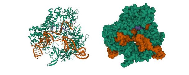
DNA polymerase beta (POLB) complexed with seven base pairs of DNA and deoxythymidine triphosphate (dTTP), 3D cartoon model in two perpendicular projections, white background

Structure of human interleukin-21, 3D cartoon model isolated with the colored elements of the secondary structure, white background

Bovine mitochondrial ATP synthase, 3D Gaussian surface model, white background, isolated

Chemical formula, structural formula and 3D ball-and-stick model of a hormone and neurotransmitter norepinephrine (noradrenaline), white background

Chemical formula, structural formula and 3D ball-and-stick model of a hormone and neurotransmitter epinephrine (adrenaline), white background

Chemical formula, skeletal formula and 3D ball-and-stick model of lignocaine (lidocaine), a local anesthetic of the amino amide type, white background

Chemical formula, skeletal formula and 3D ball-and-stick model of benzocaine, a local anesthetic of the ester type, white background
Structure of the active adrenomedullin 1 receptor G protein complex with adrenomedullin peptide (light brown), membrane shown. 3D cartoon Gaussion surface models, PDB 6uun, white background.

Crystal structure of human matrix metalloproteinase MMP9 (gelatinase B). 3D cartoon model, secondary structure color scheme, PDB 1l6j, white background

An aerial view of the Harrat Kishb volcanic field, Makkah Province, Saudi Arabia
Structure of Streptococcus pyogenes Cas9 in complex with guide RNA (blue) and target DNA (brown). 3D cartoon and Gaussian surface models, chain entity color scheme, PDB 4oo8, white background

Cas1(green)-Cas2(brown)-prespacer binary complex. 3D cartoon and Gaussian surface models, chain entity color scheme, PDB 5xvn, white background

Cas1-Cas2-Csn2-DNA complex from the Type II-A CRISPR-Cas system. 3D cartoon and Gaussian surface models, chain entity color scheme, PDB 6qxf, white background
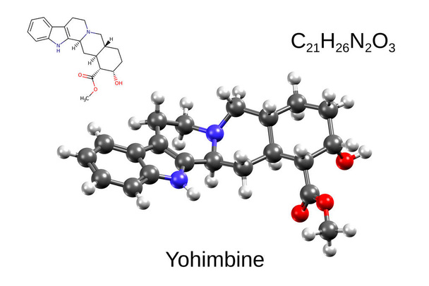
Chemical formula, skeletal formula, and 3D ball-and-stick model of yohimbine, a drug for erectile dysfunction, white background

Crystal structure of human CD9, 3D cartoon and Gaussian surface models, white background

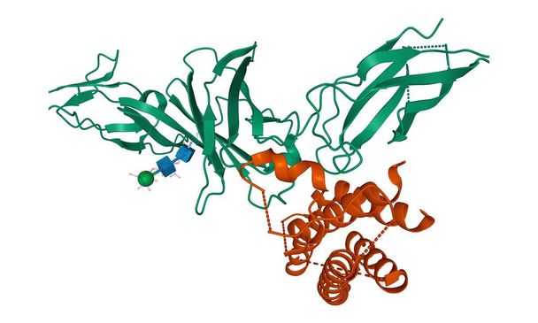
Crystal structure of human epidermal growth factor dimer. 3D cartoon and Gaussian surface models, chain id color scheme, PDB 1jl9, white background

Chemical formula, structural formula and 3D ball-and-stick model of antibiotic chloramphenicol, white background

Chemical formula, structural formula and 3D ball-and-stick model of macrolide antibiotic erythromycin

Crystal structure of human polynucleotide phosphorylase, 3D cartoon and Gaussian surface models, PDB 3u1k, white background.

Structure of Taq DNA polymenrase, 3D cartoon model, secondary structure color scheme, based on PDB 1taq, white background

Helicobacter pylori Vacuolating Cytotoxin A Oligomeric Assembly 1, 3D cartoon and Gaussian surface models, chain id color scheme, PDB 6nyf, white background

Structure of human insulin-degrading enzyme (blue) in complex with insulin (green-yellow). 3D cartoon model, PDB 2wby, white background.

Structure of human octameric PAICS, 3D cartoon and Gaussian surface model, white background.

Structure of human CD1a (green) in complex with beta-2-microglobulin (brown), 3D cartoon model isolated, white background

Crystal structure of the human T-cell co-receptor CD8 homodimer, 3D cartoon model, white background

Structure of human interleukin-23 heterodimer, 3D cartoon and Gaussian surface models isolated, white background
Crystal structure of CD5_DIII with the differently colored secondary structure elements, 3D cartoon model, white background

Structure of human CD1e, 3D cartoon model isolated, white background

CD1b (green) in complex with beta-2-microglobulin (brown) and GM2 ganglioside (pink), 3D cartoon model, white background

Structure of sodiumpotassium adenosine triphosphatase, also known as the sodiumpotassium pump, 3D cartoon model, white background

Structure of the alpha1beta1gamma2S tri-heteromeric GABAA receptor in complex with GABA (spacefill), 3D cartoon model with reduced opacity, white background

Crystal structure of human CD33, 3D cartoon model of homodimer with differently colored chains, white background

Crystal structure of human CD33, 3D surface model of homodimer with differently colored chains, white background
Structure of human interleukin-12 heterodimer, 3D cartoon model isolated, white background

Crystal structure of the human CD40 ligand homotrimer, 3D cartoon model, white background

Structure of the trimeric globular domain of adiponectin, 3D cartoon model isolated, white background

A 3D cartoon model of the human CD94 C-type lectin receptor, white background
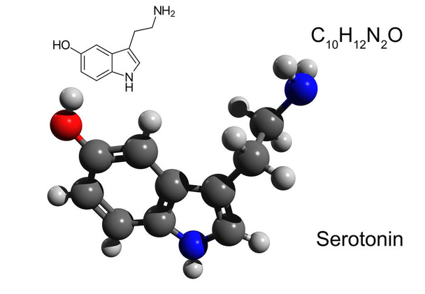
Chemical formula, structural formula and 3D ball-and-stick model of a neurotransmitter serotonin, white background

Structure of novel coronavirus spike receptor-binding domain (pink) complexed with its receptor ACE2 (blue), white background

Structure of the SARS-CoV-2 spike glycoprotein, surface model, black background, 3D illustration isolated
Structure of the Apaf-1 apoptosome with cytochrome C shown, 3D cartoon model, black background

Chemical formula, structural formula and 3D ball-and-stick model of the anticancer drug vemurafenib, white background

Structure of the SARS-CoV-2 spike glycoprotein, surface model, white background, 3D illustration isolated

Chemical formula, structural formula and 3D ball-and-stick model of antiviral drug nevirapine, white background

Structure of human respiratory complex III, 3D cartoon and Gaussian surface models, chain id color scheme, based on PDB 5xte, white background

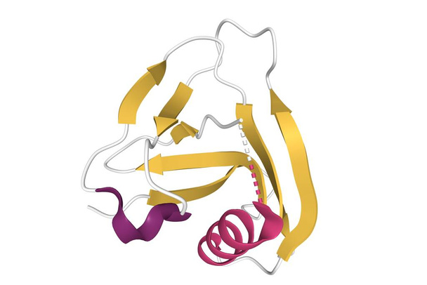
Structure of human alpha-defensin-5, 3D cartoon model isolated with differently colored secondary structure elements, white background

Human TFIID bound to promoter DNA and TFIIA, 3D cartoon model, black background

The sport utilities vehicles near the Cathedral Stone, an isolated natural sandstone formation and popular tourist attraction in the desert near Riyadh

Chemical formula, skeletal formula and 3D ball-and-stick model of a non-steroidal anti-inflammatory drug clonixin, white background

Chemical formula, structural formula and 3D ball-and-stick model of azapropazone, a nonsteroidal anti-inflammatory drug, white background

Chemical formula, structural formula and 3D ball-and-stick model of lornoxicam, also known as chlortenoxicam, a nonsteroidal anti-inflammatory drug (NSAID) of the oxicam class, white background

Chemical formula, structural formula, and 2D ball-and-stick model of acetyl-CoA, white background

Chemical formula, structural formula and 3D ball-and-stick model of DAPI, a fluorescent stain, white background

Chemical formula, skeletal formula, and 3D ball-and-stick model of chemotherapeutic drug amsacrine, white background

G2-mitotic-specific cyclin B1 (pink), cyclin-dependent kinase 1 (blue) and cyclin-dependent kinase regulatory subunit 2 (green) with Flavopiridol. 3D cartoon and Gaussian surface models, PDB 6gu2, white background

Crystal structure of CDK4 (pink) in complex with cyclin D1 (blue) and P27 inhibitor (green). 3D cartoon and Gaussian surface models, PDB 6p8e, white background

Structure of cyclin-dependent kinase 2 (CDK2, blue) in complex with cyclin E (brown). 3D cartoon and Gaussian surface models, PDB 1w98, white background

Structure of cyclin-dependent kinase CDK9 (green) in complex with cyclin T (brown) and a 2-amino-4-heteroaryl- pyrimidine inhibitor. 3D cartoon and Gaussian surface models, PDB 4bci, white background

Chemical formula, structural formula and 3D ball-and-stick model of antibiotic clindamycin, white background

Crystal structure of the retinoblastoma tumor suppressor protein (AB domain, green and brown) bound to E2F peptide (red), 3D cartoon and Gaussian surface models, chain id color scheme, PDB 1o9k

Cryo-EM structure of SARS-CoV-2 ORF3a, 3D cartoon and Gaussian surface models, based on PDB 7kjr, white background

Structure of mosquito-larvicidal toxin Cry4Ba from Bacillus thuringiensis, 3D cartoon and Gaussian surface models, chain instance color scheme, PDB 1w99, white background

Helicobacter pylori Vacuolating Cytotoxin A Oligomeric Assembly 2a. 3D Gaussian surface model in two purpendicular projections, chain id color scheme, PDB 6nyg, white background