


Filters

Stock royalty-free photos and images of Biopsie
Discover unlimited high resolution images of Biopsie and stock visuals for commercial use.

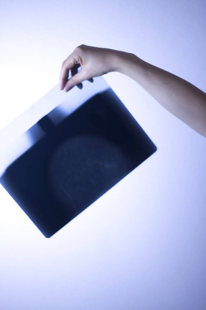
Scientist hold slide in left hand and write something down on piece of white paper by right hand in laboratory with vignetting effect
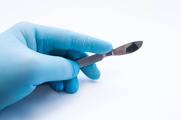
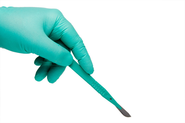
Hand holding scalpel. Palm of surgeon dressed in blue glove holding scalpel. Concept photo for surgeries, procedures, treatment, plastic surgery operation, work of surgical departments and hospitals

Background concept wordcloud illustration of prostate cancer glowing light

Woman checking suspicious mall on her skin
Human anatomy exploded view icon, 3D illustration

Human anatomy exploded view, 3D illustration

3d security forces people illustration. Scientific police analyzing forensic evidence with a microscope. Isolated white background.
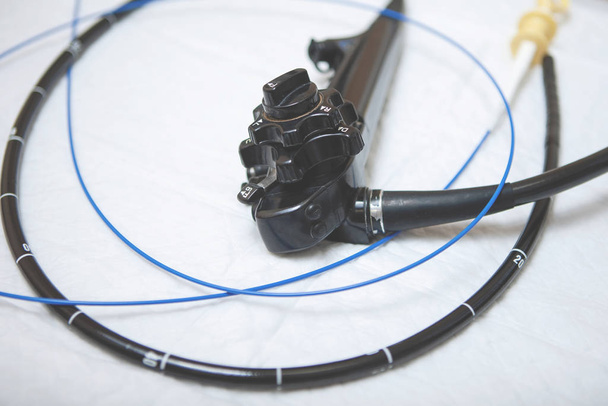
Gastroscopy, endoscopy. Apparatus for cavity research: fibrogastroduodenoscopy, a study of the stomach

3d rendering endoscope remove colonic polyp with wire loop

Background concept wordcloud illustration of biopsy glowing light

Writing word PROSTATE CANCER with marker on gradient background made in 2d software

Handwritten Diagnosis Thyroid Cancer in the Disease Extract. Medicaments Composition of Heap of Pills, Blister of Pills and Bottle of Tablets. 3D Render.
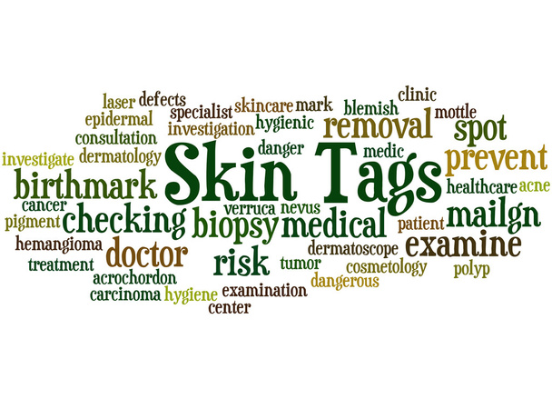
Skin Tags, word cloud concept on white background.

Skin Tags, word cloud concept on black background.

Animal tissue samples under the microscope in Lab.
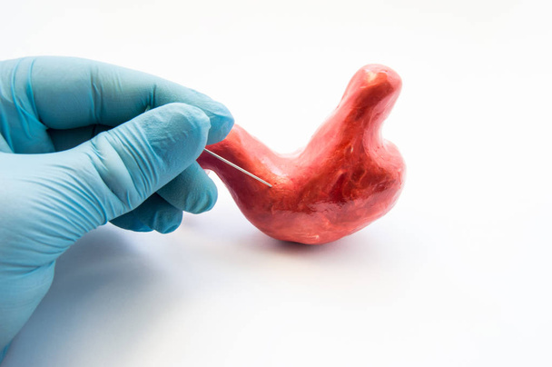
Concept of stomach puncture or gastrointestinal perforation. Hand of surgeon pierces wall of model of human stomach for therapeutic purposes or for biopsy tissue analysis by histology or cytology
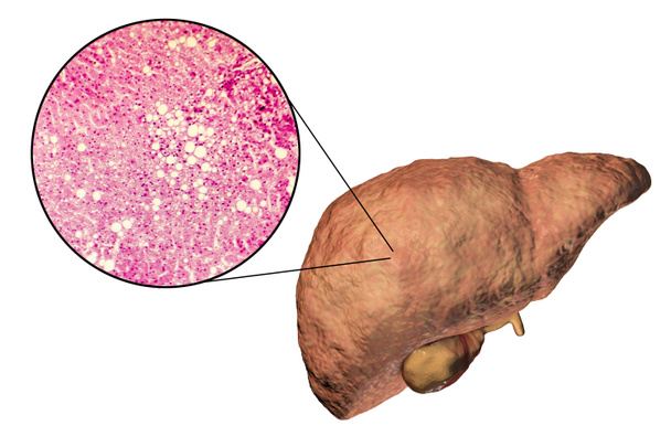
Fatty liver, liver steatosis, 3D illustration and photomicrograph showing large vacuoles of triglyceride fat accumulated inside liver cells, it occurs in alcohol overuse, under action of toxins
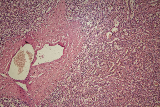
Histopathology of prostate gland hyperplasia, light micrograph, photo under microscope
Thyroid cancer cells, 3D illustration

All products lactose free and gluten free

Woman looking suspicious mall on her skin
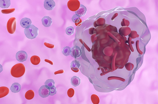
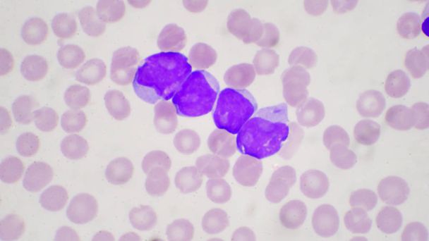
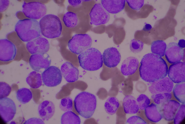
Cells of a human spleen with chronic myelogenous leukemia, under the microscope.

Cancer Diagnosis. Medical Concept with Composition of Medicaments - Red Pills, Injections and Syringe. Selective Focus. 3D Render
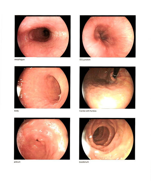
Gastrointestinal endoscopic examination,Finding esophagus,EG junction,body,cardia with fundus,antrum,duodenum normal contains excessive noise, film grain, compression artifacts when full solution.

Scientific background with modern histopathology tools: stained microscopic slides of patient tissue, fixed tissue samples and a microscope. Space for your text. This image is toned.
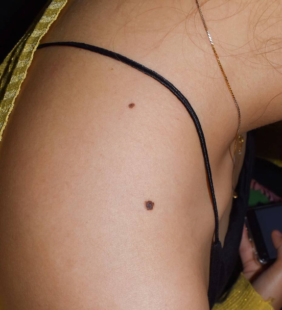
Junctional nevus or double moles at shoulder of Southeast Asian, Myanmar young woman. It is found at border between epidermis and dermis layers of skin. These moles may be colored and slightly raised.
Gastroenterology medical concept image with icons and doctors on background

Doctor examining woman skin for melanoma

Student girl looking in a microscope, science laboratory concept. Portrait of beautiful young woman in a laboratory sitting on her workplace.

Lactose free and gluten free

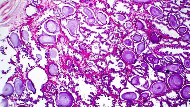
Prostate hyperplasia. Photomicrograph showing dilated glands, enfolding of the glandular epithelial cells forming papillary projections into the gland lumen, cystic dilatation
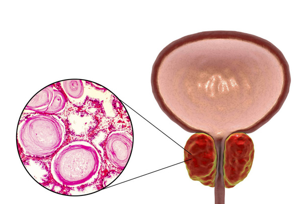
Benign prostatic hyperplasia, 3D illustration showing enlarged prostate gland and photomicrograph showing dilated glands, enfolding of the glandular epithelial cells, cystic dilatation
A medical scalpel after a surgical procedure.
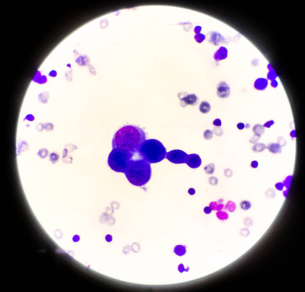
Blast cell in leukemia pateins in blood smear cbc.

Young woman doctor's hands close up preparing for an ultrasound device scan.

Team of surgeons doing an operation. Sterile operating room. Kidney surgery in a small patient. Surgical procedures Diagnostic operation

Laboratory equipment composition. Science concept.

Intestinal polyps closeup - white skin -- 3D Rendering

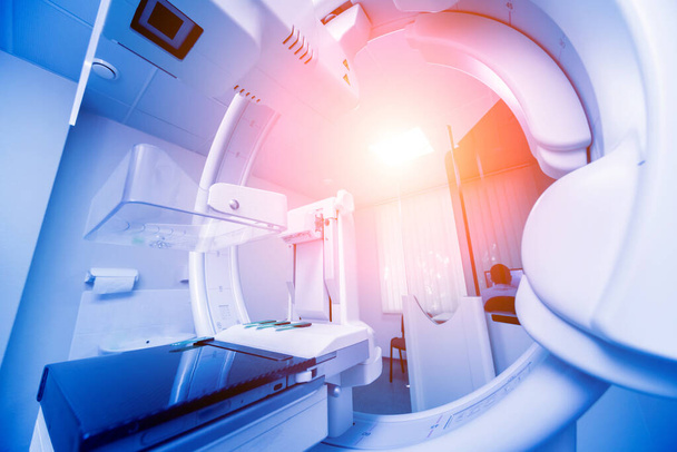
3d white radiologist looking a radiography, isolated white background, 3d image
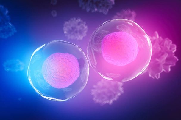
Body cells under a microscope. A good illustration as a representation of research of stem cells, cellular therapy and regeneration and many other concepts. 3d illustration
Squamous epithelial cells under microscope view for education histology. Human tissue.

Primary myelofibrosis (PMF) cells in blood flow - closeup view 3d illustration
Anesthesiology medical concept image with icons and doctors on background
Oncology medical concept image with icons and doctors on background
Diagnosis medical concept image with icons and doctors on background

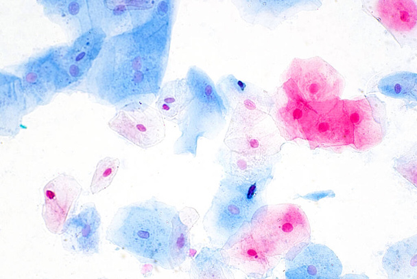
Squamous epithelial cells of human cervix under the microscope view. Pap smear test is a procedure to test for cervical cancer in women.
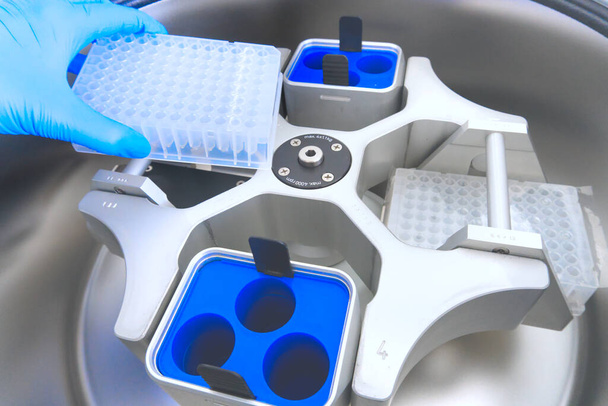
Scientist puts the test tubes in centrifuge. Automation in the clinical laboratory. Pipetting robot laboratory. Research and science background.
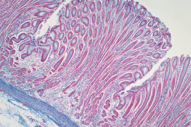
Colon biopsy from the colonoscopy. The pathology report describes normal colonic mucosa fragment with colic glands. Haematoxylin and eosin staining technique slide.
Auer rods (or Auer bodies) in acute Hypergranular Promyelocytic Leukemia (APL) - 3d illustration closeup view
CAR T cell therapy in Multiple myeloma (MM) - isometric view 3d illustration

Gastroscopy, endoscopy. Apparatus for cavity research: fibrogastroduodenoscopy, a study of the stomach
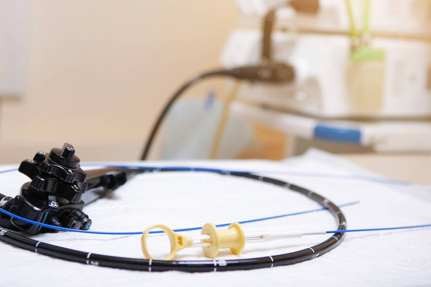
Gastroscopy, endoscopy. Apparatus for cavity research: fibrogastroduodenoscopy, a study of the stomach
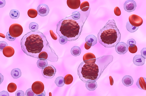
Primary myelofibrosis (PMF) cells in blood flow - isometric view 3d illustration
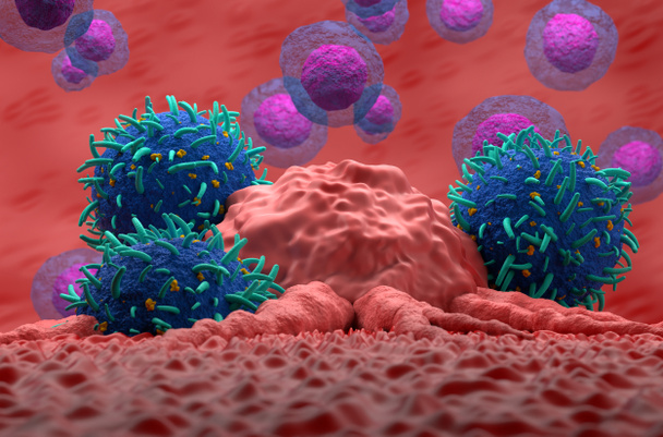
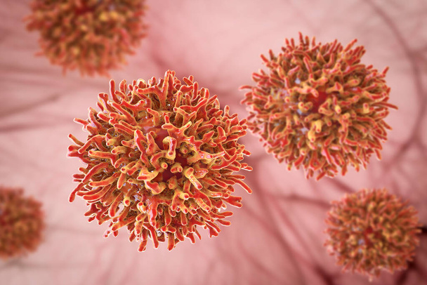
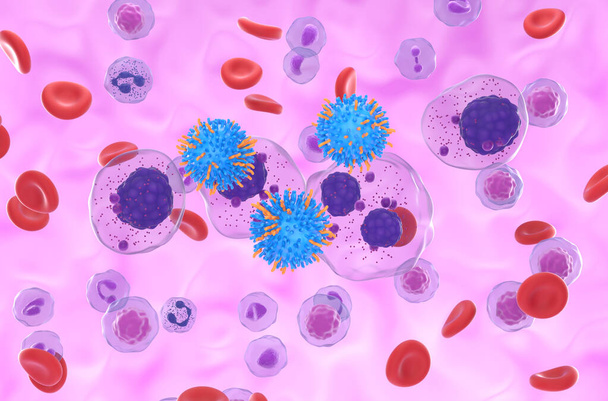
T-cells attack cancer tumor 3D render illustration closeup

Stomach ulcer - high degree of detail -- 3D Rendering
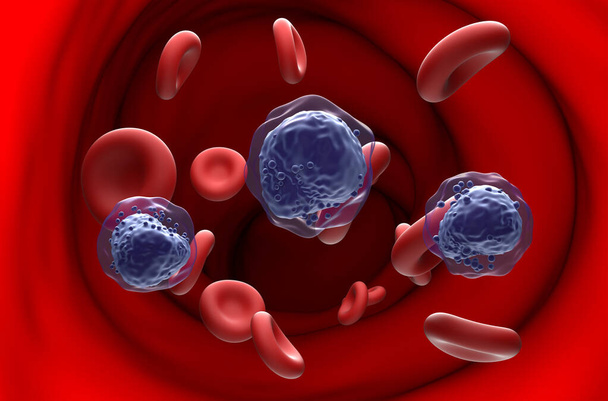
Acute myeloid leukemia (AML) cells in blood flow - section view 3d illustration

Intestine with Morbus Crohn. Illustration

Precision Oncology and Targeted Oncology Therapy - Area of medicine that Uses Personalized and Genomic-based Approaches to Diagnose and Treat Cancer
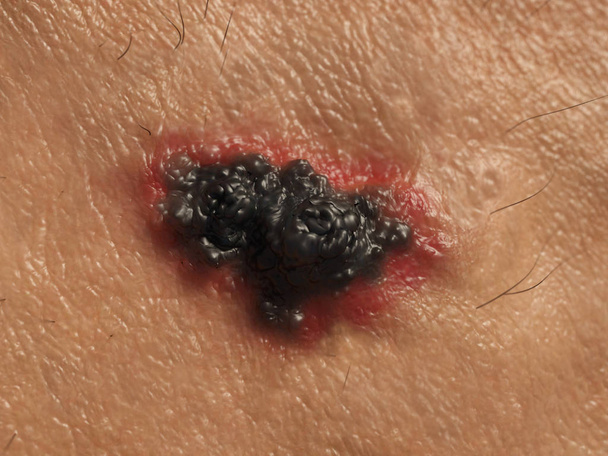
Human Skin cancer. Illustration

Tumor cell in the moment that divides, 3d illustration

Prostate cancer stages. ancerous cells, malignant tumor compresses urethra. Pathological disruption, genital reproductive system anatomy, bladder Oncological or Urological Disease, bph, 3d render

Areolar connective tissue under the microscope view. Histological for human physiology.

Wooden Blocks with the text: Cancer

Laboratory interior. Laboratory equipment glass beakers and microscope. Science concept.

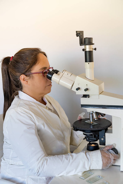
Woman in a laboratory microscope with microscope slide in hand.toned image
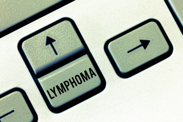
Handwriting text Lymphoma. Concept meaning Cancer that begins in infection fighting cells of the immune system.

Conceptual hand writing showing Melanoma. Business photo text A malignant tumor associated with skin cancer Benign moles.

Writing note showing Melanoma. Business photo showcasing A malignant tumor associated with skin cancer Benign moles.

Conceptual hand writing showing Lymphoma. Business photo text Cancer that begins in infection fighting cells of the immune system.

Writing note showing Melanoma. Business photo showcasing A malignant tumor associated with skin cancer Benign moles.

Text sign showing Melanoma. Conceptual photo A malignant tumor associated with skin cancer Benign moles.

Writing note showing Lymphoma. Business photo showcasing Cancer that begins in infection fighting cells of the immune system.

Microscope with lab glassware science laboratory research, Science for Health Care Concept
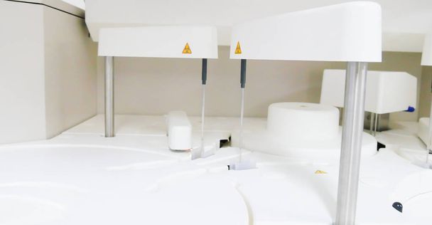
Close up on automate chemistry laboratory equipment in white tone.
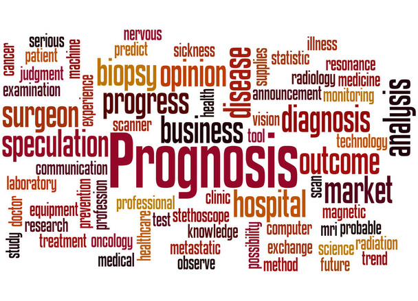
Prognosis, word cloud concept on white background.
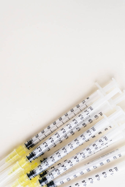
Fine syringes for use in medicine
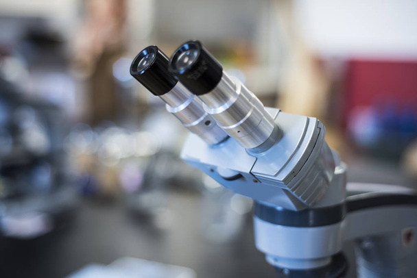
Scientific microscope close up with blurry background

Asian/Indian male scientist or doctor or science student experimenting with microscope and chemicals, laptop and smartphone in a lab

Asian/Indian male scientist or doctor or science student experimenting with microscope and chemicals, laptop and smartphone in a lab
Gastroenterology medical concept image with icons and doctors on background
Insurance medical concept image with icons and doctors on background
General surgery medical concept image with icons and doctors on background

Word writing text Cervical Cancer. Business photo showcasing type of cancer that occurs in the cells of the cervix Elements of this image furnished by NASA

Man in protective suit, medical mask and rubber gloves for protect from bacteria and virus. Quarantine, world pandemic, COVID-19, coronavirus, infection.
Mammography test at the hospital. Medical equipment. Background

Histologist with a microscope and a tray of microscopic slides on a white background

Young female scientist or tech writes down results of microsopic observations in research lab
Medical hospital mammography x-ray breast cancer mammogram scan.

Image of scientist using microscope in medical laboratory

Doctor operating ultrasound scanner with green screen. Rear view shot of a male practitioner using ultrasound scanner with green mock up screen. Physician smiling to the camera

Microscope photo of a section through cells of a frog liver.
Blast cell in leukemia pateins in blood smear cbc.
Abnormal cells in pleural fluid wright staining.
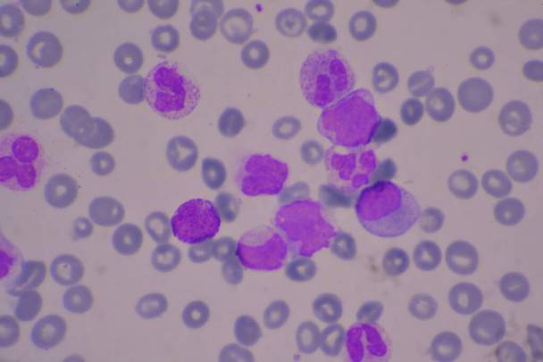
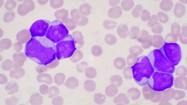
Immature and mature white blood cells.Segmented neutrophil,blast cells myelocyte,metamyelocyte,Band form.
Immature and mature white blood cells.Segmented neutrophil,blast cells myelocyte,metamyelocyte,Band form.

Doctor keeps a card with the name of the diagnosis - oncology. Selective focus. Medical concept
Scientist with her white apron sitting on a chair with her red glasses in her laboratory observing a sample through a microscope

Handwriting text Cervical Cancer, Business overview type of cancer that occurs in the cells of the cervix Questioning Uncertain Thoughts, Discussing Unresolve Problems

Text caption presenting Cervical Cancer, Business overview type of cancer that occurs in the cells of the cervix Colorful Idea Presentation Displaying Fresh Thoughts Sending Message

Conceptual display Cervical Cancer, Business concept type of cancer that occurs in the cells of the cervix