


フィルター

Arterial circulationロイヤリティフリーの写真と画像をストックする
Arterial circulation無制限の高解像度画像と商用利用用のストックビジュアルをご覧ください。

レントゲンは胸部の左側にあります。ハートぺース システム。支援システムの心臓のポンプのとおり.

胸の左側のレントゲン写真。埋め込まれたペースメーカーシステムが付いている拡大された心臓。以下は心臓アシストシステムのポンプです.

腹部大動脈瘤です。腹部の動脈循環系.健康な腹部大動脈と動脈瘤の腹部大動脈.黒を基調とした平面的なイラスト

腹部大動脈瘤です。腹部の動脈循環系.健康な腹部大動脈と動脈瘤の腹部大動脈.黒を基調とした平面的なイラスト

オーケストラ・アンソニー。動脈構造図図

心臓は筋肉器官、長い循環系の血管を介してポンプ血影、3 d レンダリング

腹部大動脈瘤です。腹部の動脈循環系.健康な腹部大動脈と動脈瘤の腹部大動脈.黒を基調とした平面的なイラスト
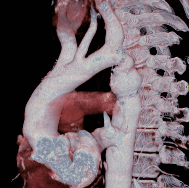
Aortic coarctation. Here, a considerable narrowing of the aorta is visible, just past the point where the aorta and the subclavian artery meet, causing arterial hypertension. 3D CT scan.

The arterial canal carries blood from the pulmonary artery to the aorta during fetal life and normally closes within hours after birth. In certain cases, however, the canal remains open increasing blood flow into the pulmonary artery, causing increas

解剖学的神経学のイラスト。胸部動脈性動脈瘤.

脳の解剖学的構造と脊髄のイラスト

塞栓症 - 医療の概念と黒い黒板。医療コンセプト: テキストと緑の聴診器に塞栓症 - 手で黒い黒板が描画されます。平面図です。3 d レンダリング.

心臓発作のコンセプト

静脈瘤に苦しむ人々は、しばしば遅すぎる病理学者に来る-目に見える兆候がすでに足に現れた瞬間:腫れ,皮膚の構造の変化,潰瘍,腫れ静脈.
高血圧の看板を止める - 開いた手を上げ、白い背景に隔離

それで書かれたテキストの高圧と聴診器で医師

人間の脳の解剖図

診療所の患者の腕に血圧と脈拍を取る医師

Pacemaker isolated on a white background. Heart battery. Close-up of cardiac pacemaker on electrocardiography.

Fla source file available - The heart is a muscular organ about the size of a fist, located just behind and slightly left of the breastbone.

聴診器やスタジオ写真で青の背景に医薬機器

Pacemaker isolated on a white background. Heart battery. Close-up of cardiac pacemaker on electrocardiography.

アテローム性動脈。動脈が部分的に塞がってる。動脈壁のプラーク

3Dイラスト- Narrowed Carotid Artery

The Nephron function or Reabsorption 3D rendering